Monthly Archives

September 2015

HIV

By Applying for disability benefits when you have:

Can I Get Social Security Disability Benefits for HIV / AIDS?

  • How Does the Social Security Administration Decide if I Qualify for Disability Benefits for HIV / AIDS?
  • About HIV Infection and Disability
  • Winning Social Security Disability Benefits for HIV / AIDS by Meeting a Listing
  • Residual Functional Capacity Assessment for HIV / AIDS
  • Getting Your Doctor’s Medical Opinion About What You Can Still Do

How Does the Social Security Administration Decide if I Qualify for Disability Benefits for HIV / AIDS?

If you are infected by HIV, Social Security disability benefits may be available. To determine whether you are disabled by HIV / AIDS, the Social Security Administration first considers whether your HIV / AIDS is severe enough to meet or equal a listing at Step 3 of the Sequential Evaluation Process. See Winning Social Security Disability Benefits for HIV Infection by Meeting a Listing. If you meet or equal a listing because of HIV, you are considered disabled. If your HIV / AIDS is not severe enough to equal or meet a listing, the Social Security Administration must assess your residual functional capacity (RFC) (the work you can still do, despite your HIV), to determine whether you qualify for disability benefits at Step 4 and Step 5 of the Sequential Evaluation Process. See Residual Functional Capacity Assessment for HIV Infection.

About HIV Infection and Disability

What Is HIV?

Human immunodeficiency virus (HIV) causes acquired immunodeficiency syndrome (AIDS). The most common type of HIV in the U.S. is HIV-1. Another type of HIV, HIV-2, is more common in Africa but is spreading to other countries. HIV-1 and HIV-2 have a 40% similarity in DNA. Current HIV testing can detect either type, but HIV-2 cases are still relatively rare in the U.S.

Origin of HIV

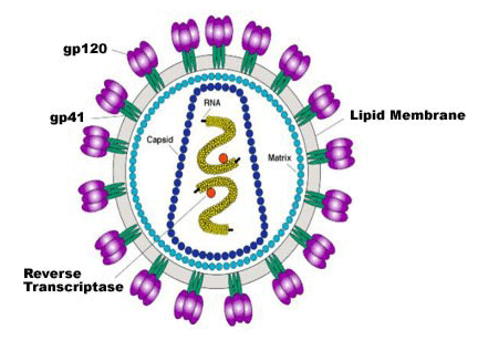
Sophisticated genetic research shows that HIV-1 (see Figure 1 below) originated in Africa and was transmitted from monkeys to the human population in about 1908, existing at low levels until the middle of the 20th century when the development of population centers facilitated spread.

While it was previously thought that monkeys had immunity from HIV because of its presence in them for perhaps millions of years, it is now known that monkey infections have not existed much longer than human infections. The sootey mangabey monkey first caught simian immunodeficiency virus (SIV) in around 1808 and it jumped into humans to become HIV-2 in about 1933. It is still not known why monkeys carrying these viruses suffer no ill effect, while they are so devastating to humans.
The HIV-1 Virus

Figure 1: The HIV-1 virus.

Infection Rates

As of 2008, the global infection rate is estimate to be about 33.2 million people with a cumulative death toll to date of about 25 million people. In the U.S.it is estimated that there are about 1.1 million HIV infected people and about 25% of these don’t know they are infected. These carriers nevertheless remain potentially infectious to other people.

Transmission

The majority of cases are now thought to occur by heterosexual transmission. New HIV infections are still growing in the U.S. at the rate of about 40,000 to 50,000 per year. HIV infection is also seen in IV drug users, homosexual and bisexual men, and others who engage in high-risk sexual activity such as prostitution and unprotected sex.

There is no risk of infection from casual contact, as living in the same household. Although HIV exists in saliva, transmission by kissing has been demonstrated in only one case when both individuals had gum disease so that there was exposure to blood. HIV is present in an infected mother’s milk.

Testing of the blood supply prior to transfusion is carried out for both HIV-1 and HIV-2.

Effects of HIV Infection

Infection with HIV is not AIDS and may produce no symptoms. However, eventually HIV suppresses the patient’s immune system by destroying the CD4 helper T lymphocytes. This opens the door to easy development of cancers, bacterial infections, viral infections, fungal infections, protozoan infections, parasitic infections, and general debilitation. Additionally, HIV itself may damage organs such as the brain.

When such severe consequences of having immune suppression as result of HIV infection appear, then the patient has developed AIDS. The listing for HIV lays out the broad areas of impairment that can potentially be manifested in AIDS. If the listing is satisfied, the claimant has AIDS. See Winning Social Security Disability Benefits for HIV / AIDS by Meeting a Listing.

The average normal CD4 lymphocyte count is 500 to 1300 per mm3 of blood (average, 800 per mm3). Opportunistic infections, cancer and other manifestations of AIDS typically appear when the CD4 count falls to 200/mm3 or less.

Treatment

When to Begin

It remains controversial at just what CD4 count therapy should start, although everyone agrees that people with 200/mm3 or less should be treated. The issue is controversial because starting treatment with CD4 counts high enough that the patient has no clinical disease subjects the patient to the increased danger of creating drug-resistant HIV and the toxicity of the drugs. On the other hand, starting treatment with too low of a CD4 count imperils recovery of the immune system.

Patients have a life-expectancy of at least 7 years on drug regimens that were started with CD4 counts above 500/mm3 and at least 30,000 copies of HIV RNA in the blood, while patients starting treatment at lower CD4 counts averaging 87/mm3 have done much more poorly with a life expectancy of less than 3 years.

In 2001 the U.S. Department of Health and Human Services (HHS) recommended that treatment be started at a CD4 count of 350/mm3 or a viral load higher than 55,000 copies of HIV RNA per mm3 of blood. Also, it is agreed that those few patients who are detected within 6 months of infection should be treated in an attempt to save immune functions that will otherwise be irretrievably lost. A large-scale, multi-year clinical trial at a cost of $121 million by the National Institute of Allergy and Infectious Diseases started in 2002. It is expected to involve 6000 patients and take 9 years. Not all experts are enthusiastic that even this expensive trial will answer needed questions about just when therapy should start. For one thing, better control of drug toxicity could affect clinical decisions to start earlier treatment.

In 2006, the International AIDS Society—USA released its newest guidelines for antiretroviral therapy, and continues to recommend that treatment in both symptomatic and asymptomatic individuals start when CD4 counts fall below 350 cells/microliter and before they reach 200 cell/microliter.

No Cure

Regardless of the treatment given or when it is started, therapy cannot cure HIV infection or AIDS. Patients who have been treated for years with potent drugs to the extent that there is no detectable virus in the bloodstream will relapse into active infection if medication is stopped. HIV can attach itself to the chromosomes of white blood cells and pass itself along to new cells when the old ones die. In this situation, the virus does not replicate and therefore is not susceptible to drugs. There is also evidence that the virus can lie dormant in other tissues besides white cells. Therefore, medication must be continued indefinitely.

Drugs for Treating HIV

Antiretroviral medications inhibit the reproduction of the HIV virus. There are six classes: non-nucleoside reverse transcriptase inhibitors, nucleoside reverse transcriptase inhibitors, protease inhibitors, entry inhibitors, fusions inhibitors, and integrase inhibitors. They each work in a different way. Non-nucloeside reverse transcriptase inhibitors work by inhibiting the reverse transcriptase enzyme that HIV needs to change its RNA to DNA in order to infect the nucleus of the cell it has invaded. Nucleoside reverse transcriptase inhibitors prevent reproduction of the virus by providing it with faulty components that it needs to make copies of itself. Protease inhibitors interfere with replication of the virus after it has infected the host cell’s nucleus. Entry inhibitors and fusion inhibitors prevent HIV from entering cells. Integrase inhibitors inhibit a protein that HIV uses to insert its genetic material into the genetic material of an infected cell.

Highly active antiretroviral therapy (HAART), the current recommended treatment for HIV, involves taking a combination of anti-HIV medications from at least two different classes.

The latest evidence shows that drug therapy has substantially lengthened the lifespan of those with HIV infection. While untreated HIV will typically result in death within 12 years, the life-expectancy of a treated HIV-infected 20-year-old has increased from 56.1 years in 2005 to 69.4 years in 2005. The CD4 cell count is also important. A 20-year-old with a CD4 count of less than 100/microliter has a life expectancy of 32.4 additional years, while the same person started treatment CD4 count over 200/microliter can expect 50.4 more years of life.

Side Effects of Treatment

It is possible for some people to be asymptomatic on HAART once HIV loads have been suppressed and CD4 counts return to normal, provided that opportunistic infections have been controlled and there is no cancer or other chronic residual impairment. However, HIV strains are becoming increasingly resistant to drugs. Furthermore, medication side-effects can be debilitating even if the virus is kept under control.

The great majority of patients taking multiple drugs will have some type of side-effects. The most frequent side-effects are nausea, vomiting, insomnia, fatigue, malaise, and headache. Other possible side-effects include anemia, pancreatitits, peripheral neuropathy, general decrease in white cells (pancytopenia), cough, diarrhea, sore throat (pharyngitis), shortness of breath, muscle aches (myalgias), muscle weakness, acidosis, hepatitis, kidney stones (neophrolithiasis), rash, and fever. Side-effects will generally reverse with cessation of medication.

In addition, various impairments, such as infections and cancers, that can be associated with AIDS must also be treated. These impairments and the drugs used to treat them can add a whole additional universe of possible symptoms and complications. Therefore, each case must be carefully evaluated on an individual basis; there is no way to know ahead of time or to presume what problems will be present.

Continue to Winning Social Security Disability Benefits for HIV / AIDS by Meeting a Listing.

Lung disease

By Applying for disability benefits when you have:

Can I Get Social Security Disability Benefits for Lung Disease?

  • How Does the Social Security Administration Decide if I Qualify for Disability Benefits for Lung Disease?
  • About Lung Disease and Disability
  • Winning Social Security Disability Benefits for Lung Disease (Chronic Pulmonary Insufficiency) by Meeting a Listing
  • Residual Functional Capacity Assessment for Lung Disease
  • Getting Your Doctor’s Medical Opinion About What You Can Still Do

How Does the Social Security Administration Decide if I Qualify for Disability Benefits for Lung Disease?

If you have lung disease, Social Security disability benefits may be available. To determine whether you are disabled by your breathing problems, the Social Security Administration first considers whether your lung disease is severe enough to meet or equal a listing at Step 3 of the Sequential Evaluation Process. See Winning Social Security Disability Benefits for Lung Disease by Meeting a Listing. If you meet or equal a listing because of lung disease, you are considered disabled. If your breathing problems are not severe enough to equal or meet a listing, Social Security Administration must assess your residual functional capacity (RFC) (the work you can still do, despite your lung disease), to determine whether you qualify for benefits at Step 4 and Step 5 of the Sequential Evaluation Process. See Residual Functional Capacity Assessment for Lung Disease.

About Lung Disease and Disability

How Respiration Occurs

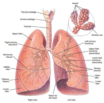
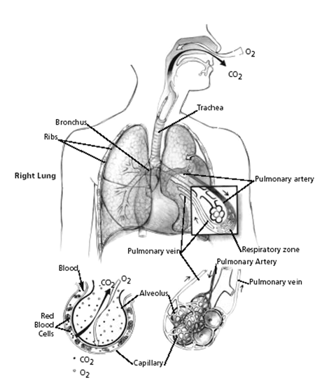
Red blood cells must be brought as closely as possible to the air we breathe, so that hemoglobin in the cells can give up waste carbon dioxide from cellular metabolism and take on oxygen. To accomplish this, the lungs have millions of tiny air sacs (alveoli) with very thin walls (alveolar membranes) containing microscopic blood vessels (capillaries) (see Figure 1 below). This anatomy of the lungs allows exposure of a large surface area of the blood to the air. Oxygen (O2) and carbon dioxide (CO2) gases diffuse (move) across the one cell thick alveolar membrane in opposite directions with the oxygen entering the blood and the carbon dioxide leaving it. This process is known as gas exchange.

Bronchi and lungs

Figure 1: Bronchi and lungs.

The gas exchange part of the lungs is known as the lung parenchyma (see Figure 2 below). Air is delivered to the parenchyma of the lungs through the bronchial tree — a repetitively branching tubular system for air conduction. It consists of the trachea, from which arises a right and left main (primary) bronchus to the right and left lungs. Smaller bronchi branch from the main bronchus of the right or left lung, then to smaller bronchi to the various lobes of the lungs, then to even smaller bronchi (bronchioles) that eventually reach the alveoli.

Mechanism of gas exchange

Figure 2: Mechanism of gas exchange.

How Respiration Is Impaired

Many diseases can affect breathing. The most useful classification of respiratory disorders is based on the manner in which the ability of air to come into contact with the hemoglobin in red blood cells (RBCs) is disrupted.

There are only two ways in which respiration can be impaired, regardless of the exact nature of the disorder:

1. Disease that prevents adequate amounts of air from reaching the gas-exchange level of the lungs (obstructive lung disease).

2. Disease of the lung tissue itself that reduces gas exchange (restrictive lung disease).

Thus, physicians have found it broadly useful to classify respiratory disorders as obstructive or restrictive, or a combination or the two.

Chronic Obstructive Lung Disease (COPD)

Chronic obstructive pulmonary disease (COPD) is the most common type of lung disease seen by the Social Security Administration. “Obstruction” refers to the fact that air flow in and out of the lungs is impeded. The three most frequent types of COPD in adults are:

1. Emphysema.

2. Chronic bronchitis.

3. Asthma.

In emphysema, lung tissue itself is destroyed. Damaged lung tissue forms non-functional spaces that trap air, and the lungs expand. The effect of these abnormalities is to obstruct air flow to and from the lungs. Emphysema and chronic bronchitis often occur together usually in people with a history of cigarette smoking.

Chronic bronchitis can also occur from long exposure to chemical fumes associated with a particular occupation. Inflammation of the inner surface of bronchi results from exposure to irritating substances. This inflammation, along with excessive secretion of bronchial mucous glands results in bronchial narrowing. Thus, bronchial tree resistance to air flow increases—there is obstruction of air flow.

Other less common causes of COPD include cystic fibrosis and bronchopulmonary dysplasia (BPD). These disorders, as well as asthma, have separate specific listings. See Can I Get Social Security Disability Benefits for Asthma? and Can I Get Social Security Benefits for Cystic Fibrosis?

Restrictive Lung Disease

The hallmark of restrictive lung disease is loss of usable lung volume, either due to:

1. Disease of the gas exchange part of the lungs (lung parenchyma), or

2. Some disorder outside of the lungs (extrapulmonary) that prevents air from adequately ventilating normal lung parenchyma.

Examples of Parenchymal Restrictive Lung Diseases

    • Infections (bacterial, fungal, viral, parasitic). Chronic infections that damage lung tissue, such as severe bronchiectasis or advanced pulmonary TB, could result in some degree of restrictive impairment.
    • Radiation, such as used for treatment of cancer, can damage lungs. Radiation lung damage is less of a problem than in the past, because modern equipment can direct therapeutic radiation in beams precisely delivered to the tumor. To the extent that this is impossible because of the size or location of the tumor, it is possible to have fibrotic lung damage caused by radiation.
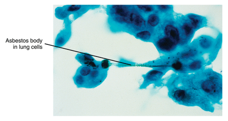
    • Inhalation of damaging substances into the lung. Pneumoconiosis is a non-specific term that refers to lung damage from inhaling small particles of some kind. Examples of pneumoconiosis include that caused by asbestos (asbestosis) (see Figure 3 below), coal dust (anthracosis), beryllium (berylliosis) silicon dust (silicosis), aluminum, iron, tin (stannosis) and talc. Inhalation of toxic chemicals—such as acidic fumes—can also damage lung tissue.

Asbestos particles in lung cells

Figure 3: Asbestos in lung cells.

  • Drug side-effects.
  • Autoimmune diseases. Autoimmune disorders such as sarcoidosis, scleroderma, systemic lupus erythematosus (SLE), rheumatoid arthritis (RA), polymyositis, ankylosing spondylitis, and mixed connective tissue disorders can cause pulmonary disease. In all of these disorders, some kind of immune system dysfunction damages the lungs. See Can I Get Social Security Disability Benefits for Lupus? and Can I Get Social Security Disability Benefits for Arthritis and Joint Damage?
  • Idiopathic diffuse interstitial pulmonary fibrosis (cryptogenic fibrosing alveolitis). This condition causes progressive scarring of the lungs on a microscopic level. This damage can result in decreased gas exchange capacity of the lungs, increasingly severe hypoxemia (lack of oxygen in the blood) and eventually death from respiratory failure. The rate of progression is highly variable, but median survival is less than 3 years. Some cases remain slowly progressive for a number of years, then are triggered by some unknown event to become rapidly more severe.
  • Other diseases. Examples of other diseases that can cause pulmonary fibrosis include alveolar proteinosis, acquired immunodeficiency syndrome (AIDS), acute respiratory distress syndrome (ARDS), amyloidosis, bone marrow transplantation, cancer, eosinophilic granuloma, eosinophilic pneumonia, lipoid pneumonia, genetic metabolic diseases caused by enzyme deficiencies (Gaucher’s disease, Niemann-Pick disease), Hermansky-Pudlak syndrome, pulmonary vasculitis, tuberous sclerosis, and neurofibromatosis. See Can I Get Social Security Disability Benefits for HIV / AIDS?

Examples of Extrapulmonary Restrictive Lung Diseases

    • Abnormal spinal curvatures. Abnormal curvatures of the spine can interfere with normal breathing movements. Scoliosis is a common spinal disorder, but will not compromise breathing until the major abnormal curve reaches about 60 degrees. Kyphosis is an abnormal curvature of the upper (thoracic) spine that causes a bent-forward or “hunched-over” posture. Kyphoscoliosis is a combination of both kyphosis and scoliosis.
    • Surgical resection. Removal of lung tissue limits the surface area available for gas exchange.
    • Spondyloarthropathies. Inflammatory disorders of the spine, such as ankylosing spondylitis, can make the spine more rigid by increased calcification of the spine and associated soft tissue ligaments. The ribs attach to the spine and their movement is impeded during expansion and relaxation of the chest during breathing. This limitation will decrease breathing capacity. See also Can I Get Social Security Disability Benefits for Back Pain?
    • Thoracoplasty. Thoracoplasty, usually involving removal of one or more ribs, distorts the normal shape of the chest. The intercostal muscles between the ribs help expand the chest during inspiration. However, mechanical distortion of the chest wall is probably more important in decreasing ability to ventilate the lung during respiratory movements of the chest.
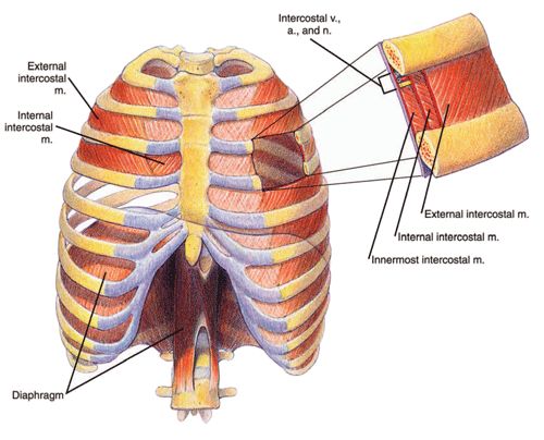
    • Obesity. Marked obesity can result in a significant reduction in ventilation capacity. Because of the weight of fat on the chest wall, the work of breathing is increased during inspiration. Furthermore, abdominal subcutaneous fat as well as intra-abdominal fat resists movement of the diaphragm—a respiratory muscle consisting of two sheets of muscle separating the chest and abdominal cavities (see Figure 4 below). The diaphragm must be able to move downward with inhalation in order to maximally expand the chest.

Chest cavity and diaphragm

Figure 4: Chest cavity and diaphragm.

  • Other important causes of restrictive respiratory impairment. Many disorders can result in weakness affecting the muscles of respiration (diaphragmatic muscles or intercostal muscles). Myasthenia gravis is an autoimmune disease that causes weakness through a biochemical interruption of the body’s ability to excite muscles, including the muscles of respiration. In fact, respiratory failure is usually the cause of death in myasthenia. There are a large number of muscle diseases (myopathies) that can affect respiration, although fairly rare in the general population.

Strokes and Breathing Problems

Strokes (cerebrovascular accidents, CVAs) are frequently adjudicated by the Social Security Administration. See Can I Get Social Security Disability Benefits After a Stroke? However, most of the time the treating source—even if a neurologist—does not consider the possibility of a breathing deficit resulting from the stroke. However, paralysis of half of the diaphragm is a definite possibility with resultant decreased ability to ventilate the lungs. Because treating sources, or consulting neurologists paid by the Social Security Administration, usually do not specifically address the possibility of diaphragmatic paralysis, SSA adjudicators are also likely to not even think of the possibility. Claimants themselves, following a stroke, may have thinking difficulties and neurological problems that occupy their attention as well as the attention of their family. The claimant cannot be counted on to mention breathing difficulty—especially since he or she may not even have symptoms in a resting state. The Social Security Administration adjudicator should ask to treating doctors, especially neurologists, regarding possible breathing deficits in post-stroke patients.

Pulmonary Function Studies

Pulmonary function study (PFS) is a general term that applies to any type of respiratory testing. The basic types of PFS are:

  • Spirometry
  • Arterial Blood Gas Study (ABGS)
  • Carbon Monoxide Diffusing Capacity (DLCO)

Shortness of breath is one of the most common allegations by claimants seeking Social Security disability benefits. The Social Security Administration pays for many PFS tests (especially spirometry) to address allegations of shortness of breath.

The results of these tests determine whether you qualify for disability benefits under a listing. See Winning Social Security Disability Benefits for Lung Disease by Meeting a Listing.

Spirometry

No pulmonary function study is more useful or more frequently performed than spirometry. Spirometry is the most important test for evaluating the severity of obstructive pulmonary disease. Spirometry requires you to inhale then exhale into a device called a spirometer. The device measures the volume of air that you can inhale and exhale and displays the result as a breathing curve on a graph called a spirogram.

If you have lung disease, but have not had a spirometry test, the Social Security Administration may arrange for you to have one. Even if you have had the test, the Social Security Administration may require you to be retested because the test must be administered in accordance with strict rules. Accurate testing must be done to assure you are treated fairly.

The test results must include the actual breathing curves. Spirometry test results in medical records often do not include the actual breathing curves, just the numerical results.

If there is no clinical evidence that you have significant lung disease and the reported spirometric values in your medical records are not significantly abnormal, the Social Security Administration will not send you for retesting. In fact, the Social Security Administration is not obligated to obtain spirometry on claimants who have no evidence in their medical records of a respiratory disorder, even if there are no spirometric values of any kind in the file.

Arterial Blood Gas Study (ABGS)

ABGS is the most important test used for the evaluation of the restrictive pulmonary disorders that involve a decrease in gas exchange—the parenchymal restrictive lung diseases. ABGS is performed on a sample of blood from the radial artery in the wrist or brachial artery in the arm, unlike most blood samples that are taken from a superficial vein just under the skin.

An ABG test checks how well your lungs are doing in moving oxygen into the blood and removing carbon dioxide from it. ABGS measures arterial oxygen pressure (PaO2), also known as oxygen tension, carbon dioxide pressure (PaCO2), and acidity (pH).

The Social Security Administration should not send you for ABGS, unless absolutely necessary to determine whether you are disabled, because it is invasive. Although complications are unusual, the person sticking a needle in the radial artery could damage the artery or other structures in the wrist; use of the brachial artery carries additional risk and should be done only when the radial artery cannot be used. The needle stick is also painful in either method.

Carbon Monoxide Diffusing Capacity (DLCO)

DLCO is a test used to evaluate of the severity of parenchymal restrictive lung diseases. It is of little value in assessing severity in COPD. The test measures how well your lungs can transfer carbon monoxide (CO) into the blood. By measuring how easily CO moves across the alveolar membrane of the lungs, doctors can deduce whether there is also a problem limiting the exchange of oxygen and carbon dioxide between blood and the atmosphere.

Two different methods are used for this test. The single-breath method is the one required by the Social Security Administration. This method requires you to take a breath of air containing a very small amount of carbon monoxide from a container while measurements are taken. (The other method, the steady-state method, requires you to breathe air containing a very small amount of carbon monoxide. The amount of carbon monoxide in the breath you exhale is then measured.)

DLCO can be abnormal in emphysema, if the disease is very advanced, because lung tissue is damaged. However, DLCO is not an accurate enough means of determining the severity of emphysema; spirometry is more appropriate. In some instances, more applicable to clinical medicine than disability determination, DLCO testing can help distinguish between emphysema and asthma, because asthma will have normal values.

Continue to Winning Social Security Disability Benefits for Lung Disease (Chronic Pulmonary Insufficiency) by Meeting a Listing.

Schizophrenia

By Applying for disability benefits when you have:

Can I Get Social Security Disability Benefits for Schizophrenia?

  • How Does the Social Security Administration Decide if I Qualify for Disability Benefits for Schizophrenia?
  • About Schizophrenia and Disability
  • Winning Social Security Disability Benefits for Schizophrenia by Meeting a Listing
  • Residual Functional Capacity Assessment for Schizophrenia
  • Getting Your Doctor’s Medical Opinion About What You Can Still Do

How Does the Social Security Administration Decide if I Qualify for Disability Benefits for Schizophrenia?

If you have schizophrenia, Social Security disability benefits may be available. To determine whether you are disabled by schizophrenia, the Social Security Administration first considers whether the schizophrenia is severe enough to meet or equal a listing at Step 3 of the Sequential Evaluation Process. See Winning Social Security Disability Benefits for Schizophrenia by Meeting a Listing. If your schizophrenia is not severe enough to equal or meet a listing, the Social Security Administration must assess your residual functional capacity (RFC) (the work you can still do, despite schizophrenia), to determine whether you qualify for disability benefits at Step 4 and Step 5 of the Sequential Evaluation Process. See Residual Functional Capacity Assessment for Schizophrenia.

About Schizophrenia and Disability

What Is Schizophrenia?

Schizophrenia is a serious mental disorder for which there is no cure. Schizophrenia affects about 1% of the adult population. There are various types of schizophrenia, i.e., disorganized, catatonic, residual, undifferentiated, and paranoid. Any of these forms of schizophrenia may entitle a person to disability benefits.

Schizophrenia appears before age 45, usually in the adolescent or young adult. The person exhibits a definite deterioration from a previous level of functioning in social, work, and personal life. Psychological abnormalities are diverse, affecting:

  • Thought.
  • Perception.
  • Emotion (affect).
  • Identity.
  • Ability to plan and carry out purposeful activity.
  • Cognition (ability to think, learn, and understand).
  • Sociability. The person may experience a tendency to withdraw from the outside world and a preoccupation with illogical ideas, fantasies, or delusions.
  • Movement. The person may exhibit a decrease, increase, or bizarreness of movements or postures.

The prevailing view of schizophrenia has been that the psychotic features are the most important and that cognitive deficits are secondary. However, the latest research suggests that the cognitive deficits, which arise from various brain abnormalities, are the more fundamental cause of mental abnormalities in schizophrenia. In fact, there is reason to think that subtle brain abnormalities appear before a child is born and gradually increase in severity.

Causes of Schizophrenia

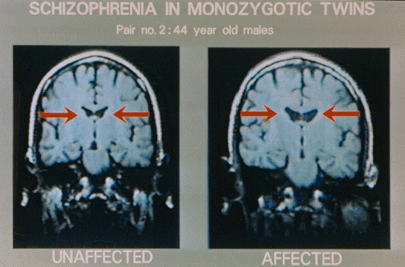
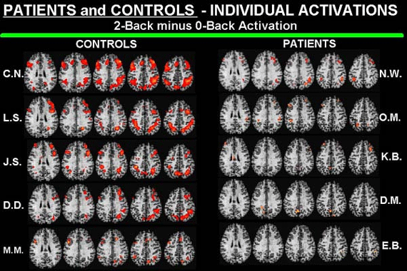
The disorder involves biochemical dysfunction in the brain, but no one can as yet describe the mechanisms involved. Numerous abnormalities are found in the brains of those with schizophrenia (see Figures 2 and 4 below).

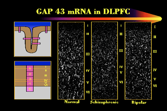
There is also a complex genetic predisposition that no one is close to understanding. It is thought that about 40% of cases of schizophrenia are inherited, and 60% occur spontaneously. Recent research suggests that schizophrenia may result from a large number of genetic abnormalities (see Figures 1 and 3 below). Consequently, there is no chance of a single cause and cure for schizophrenia.

Linkage Studies and Schizophrenia

Figure 1: A so-called “linkage study,” showing a number of places in the human genome where pieces of DNA are inherited along with risk for the illness. It shows one of each of the 23 pairs of chromosomes, and the red dots indicate regions where a piece of DNA has been shown to be inherited along with the risk for schizophrenia in certain families and certain studies.

MRI Scans and Schizophrenia

Figure 2: MRI scans of identical twins. The twin on the right has schizophrenia; the twin on the left is healthy. Even to the unprofessional eye, there are obvious differences, a systematic and consistent variation between the affected and the unaffected twin in the gross anatomy of the brain. Red arrows point to enlarged ventricles in the affected twin.

Schizophrenia and mRNA

Figure 3: Each white dot represents cells in a particular part of the brain. A patient with schizophrenia is compared to an individual with another psychiatric illness, bipolar disorder, and to a normal subject. The white dots show the turning-on of a gene that is the blueprint for a protein related to the process by which cells adapt themselves to a changing environment. I–VI represent layers of the cerebral cortex.

Schizophrenia PET Scans

Figure 4: PET scans of five normal individuals (left); each row is one person, and each image is a slice from five different levels of the person’s brain. The red areas show regions of the brain that are activated when a person performs a memory task. In PET scans of five individuals with schizophrenia (right), each row represents a different person, with comparable slices. Clearly, the patients with schizophrenia do not generate the dramatic brain activity in the circuits of the brain critical to the memory task.

Phases of Schizophrenia

Schizophrenia usually has 3 phases.

  • First, there is the prodromal phase. The duration of this phase is variable. The person shows a clear deterioration from the previous level of functioning in multiple areas of life—mental, occupational, personal, and social. The “change in personality” is often noticed by friends and relatives, as the person withdraws from usual activities, engages in less effective and increasingly strange behavior and experiences difficulty with clear and logical communication with others.
  • Next is the active or progressive phase. In this phase, overt psychotic symptoms become obvious. The impairments present in the prodromal phase become more severe. The individual increasingly withdraws. Strange behavior may become bizarre or completely disorganized, and odd perceptions and thought develop into hallucinations and delusions. Abnormalities in the form and content of thought produce further confusion and difficulty communicating with others. Transition from the prodromal to the active phase is often triggered by stress that the person cannot cope with. For example, such a stressor might be a serious physical illness or change in an important relationship with another person. Or it might be something that a normal person would consider trivial.
  • A residual phase follows the active phase. Psychotic symptoms persist to some degree, but the accompanying emotions are not as intense. (The residual phase should not be confused with the residual type of schizophrenia, so-called because an active, florid phase of the illness is missing.) Unfortunately, normal emotional responsiveness tends to be subdued also. Residual functional impairment may remain severe.

The majority of claimants seeking disability benefits for schizophrenia are in the residual phase, and in some degree of remission on neuroleptic drugs which must be taken indefinitely. However, schizophrenia almost never goes into complete remission.

Cognitive Impairment With Schizophrenia

It is very important for the Social Security Administration to pay attention to cognitive dysfunction when assessing whether a person with schizophrenia is disabled. The Social Security Administration and many psychiatrists and psychologists still emphasize the psychotic features (hallucination, delusions) of the disease. So if a claimant’s psychotic features are controlled by medication, the Social Security Administration is not likely to test memory or other cognitive functions. Fewer than 10% of patients with schizophrenia ever get a regular job or live independently. At least 90% of people with schizophrenia are apparently incapable of working on any sustained basis.

Side Effects of Schizophrenia Medications

The drugs taken to lessen the symptoms of schizophrenia can cause side effects. The severity varies from person to person. In the early phase of drug therapy, symptoms may include muscle spasms, tremors, dry mouth, drowsiness, blurry vision, and restlessness. Changing the type of medication or the dosage can help control these symptoms.

Older medications were associated with a risk of the patient developing tardive dyskinesia—a disorder characterized by involuntary movements, especially of the lips, tongue, and mouth. Newer medications, such as clozapine (Clozaril), olanzapine (Zyprexa), quetiapine (Seroquel) and resperidone (Resperdal) are associated with other risks. For example, clozapine can cause a decrease in white blood cells and greatly weaken the body’s defense against dangerous infections. Clozapine can also cause seizures, inflammation of the heart (myocarditis), low blood pressure (hypotension), and other side effects. Olanzapine (Zyprexa) can cause weight gain and Parkinsonian movement disorders. Anyone taking antipsychotic medications must be monitored by a doctor.

If the claimant is having medication side-effects, a Social Security Administration psychiatrist or other medical doctor, rather than a Social Security Administration psychologist, should evaluate at least that part of the claim.

Information from Family and Friends Is Crucial to Disability Determination

Claimants with chronic schizophrenia living with family members are most likely to be improperly denied Social Security disability benefits. It is critically important for family members or other caregivers to provide the Social Security Administration with as detailed information as possible about specific tasks the claimant can or cannot do.

Mental health clinics will often refuse to provide the Social Security Administration with clinical records that are useful in evaluating how a mental disorder has developed over time. They might simply write a letter summarizing what they think they Social Security Administration needs to know. In some instances, the Social Security Administration is forced to fall back on purchasing a consultative mental status examination in which the examining psychiatrist or psychologist has limited time to determine the details of daily functional capacity. The Social Security Administration should ask the treating psychiatrist (or psychologist) about work-related abilities for at least unskilled work and how these conclusions match with the corresponding limiting mental symptom.

If the claimant is receiving medication, information about side-effects must come from a medical doctor, because a psychologist is not competent to evaluate that matter. However, it is also important for the same kind of information and opinions to be obtained from family or other caregivers, to make sure that nothing is missed. The caregivers live with the claimant; they may have noticed important facts that can be brought to the treating psychiatrist’s attention and to the Social Security Administration.

Continue to Winning Social Security Disability Benefits for Schizophrenia by Meeting a Listing.

abogado de seguro social de Long Beach

Cómo Demuestra tus límites con dolores crónico

By Spanish Blogs

abogado de seguro social de Long BeachCómo Demuestra tus límites con dolores crónico

Ha presentado un reclamo de incapacidad con la Administración del Seguro Social (SSA) y quiere asegurarse de que saben que su dolor está limitando su vida laboral y personal? Un abogado de seguro social de Lowell puede ayudarle a comunicar cómo se siente.

Si usted está en dolor de una lesión o incapacidad, su historia medica debe reflejar eso. Un abogado del seguro social de Lowell sabe cuál es la información que usted necesita para proporcionar a SSA, que hace que la decisión sobre la cantidad, si hay algo, usted tiene derecho de recibir. Para hacer esto, es necesario comunicar la intensidad de su dolor y cómo afecta su capacidad para trabajar. Un abogado de seguro social de Lowell se reunirán al siguiente y lo presentará a la SSA:

Su historia;

  • Sus resultados médicos;
  • Las declaraciones de usted;
  • Las declaraciones de sus doctores sobre el tratamiento o los examen de los médicos; y
  • Las declaraciones de otras personas que saben acerca de cómo su dolor le afecta.

Los que toman las decisiones en la SSA también revisará su historia clínica y opiniones de los médicos que te han examinado o tratado.

Un abogado de seguro social de Lowell es consciente de que SSA considerará lo siguiente en la evaluación de su dolor:

La causa de su dolor, incluyendo lo que lo agrava ;

  • La intensidad, la frecuencia, la duración y la ubicación del dolor;
  • Su medicamento, incluyendo el tipo, la dosis, los efectos secundarios y la eficacia;
  • Cualquier tratamiento adicional además de la medicación;
  • Sus actividades diarias;
  • Otras restricciones y limitaciones funcionales; y
  • Otras medidas para aliviar el dolor que usted toma.

Un abogado del seguro social de Lowell le puede pedir los detalles los pasos para aliviar el dolor, incluyendo estar a pie de 15-20 minutos cada hora, acostado boca arriba o dormir en una tabla.

A veces el dolor es más severo que la evidencia existente sugiere, como la artritis y el dolor de espalda. Así que el que toma las decisiones no rechaza su declaración, un abogado de seguro social de Lowell hace que las decisiones en cuenta tenga credibilidad  sobre sus declaraciones.

Para obtener más información acerca de cómo probar la gravedad de su dolor, póngase en contacto con el abogado de seguro social de Lowell Gerard A. Palma al 888-295-4955.

Abogado de Lawrence Especialista en el Seguro Social Por Incapacidad

Explicación Sobre Dolor Crónico Por Un Abogado de Lawrence Especialista en el Seguro Social Por Incapacidad

By Spanish Blogs

Abogado de Lawrence Especialista en el Seguro Social Por IncapacidadExplicación Sobre Dolor Crónico Por Un Abogado de Lawrence Especialista en el Seguro Social Por Incapacidad

Una razón común por la que muchos reclamantes de incapacidad al Seguro Social dicen que no pueden trabajar es el dolor crónico. Sin embargo, un abogado de Lawrence especialista en los beneficios de incapacidad del Seguro Social le dirá que el dolor crónico no es realmente un impedimento sino que es un síntoma de un impedimento subyacente, lo que significa que no es suficiente para ganar un caso de incapacidad.

La otra desafortunada realidad acerca del dolor crónico, es que por lo general no puede ser medido por pruebas o instrumentos médicos; el único recurso que los doctores tienen es la descripción del dolor que hace el paciente. Por esta razón, un experimentado abogado especialista en incapacidad, le dirá que sus reclamos deben ser creíbles para que la Administración del Seguro Social los crea.

Preguntas A Hacer Sobre Su Situación

Hay dos preguntas pertinentes a la incapacidad cuando se trata de dolor crónico:

1) ¿Pueden usted y su abogado de Lawrence especialista en los beneficios de incapacidad del Seguro Social, proporcionar evidencia objetiva de un “impedimento médicamente determinable” que razonablemente podría causar su dolor?
2) ¿Cuán severamente su dolor le impide participar en las actividades básicas de trabajo?

Es muy importante que sus declaraciones acerca de su nivel de dolor sean consistentes con la evidencia médica y los informes suministrados por usted. Sus declaraciones acerca de su dolor, simplemente no son suficientes para demostrar que tiene un impedimento. Usted y su abogado deben proveer evidencia médica objetiva, como hallazgos de laboratorio o técnicas clínicas, de que usted tiene un impedimento físico o psicológico que probablemente sea la causa de su dolor.

Aprenda Más Con Un Abogado

Para obtener más información sobre lo que usted necesita para probar un reclamo de incapacidad, hable con un abogado de Lawrence especialista en incapacidad de Palma Law Offices, PC. Llame hoy al 888-295-4955 para pedir ayuda.

Skip to content