Category

Applying for disability benefits when you have:

Obesity Social Security Disability Claims

By Applying for disability benefits when you have:, English Blogs, Social Security Disability Library

Even though the Social Security Administration no longer considers obesity to be a listing-level severe impairment, it still remains an important element of many disability applications.  The potential effects obesity has on the musculoskeletal, respiratory, and cardiovascular body systems makes inclusion and evaluation of this condition an important part of many disability claims.

Obesity is considered a complex, chronic disease from excessive accumulation of body fat in a person.  Obesity can be a result of any of a number of factors, including genetics, nutrition, environment, behavior, and medication.

The National Institute of Health defines categories of overweight and obesity in adults based on the Body Mass Index (“BMI”).  The Body Mass Index is the ratio of a person’s weight in kilograms to the square of the person’s height in meters.  As a formula, BMI=kg/m2.  Most online calculators of a person’s body mass index will make the conversion from pounds to kilograms and inches to meters to make the process simpler.  The National Institute of Health’s guidelines state that a person is underweight at under 18.5, normal weight at 18.5-24.9, and overweight at 25-29.9.

The National Institute of Health generally defines obesity at 30 or higher and breaks it up into three levels.  Level I covers a Body Mass Index of 30.0-34.9 and Level II of 35.0-39.9.  Obesity is considered “extreme” at Level III, with a Body Mass Index of 40 and above.  People at Level III obesity are at the greatest risk for developing obesity-related impairments.  For children, there is no official criteria for making an obesity determination.  A Body Mass Index at or greater than the 95th percentile for the child’s age and gender will usually be enough to diagnose childhood obesity.  Given the different body types people have and each person’s unique physiology, the Body Mass Index is not the best indicator of a person’s fitness level.

Relevancy

While obesity is no longer considered a listing-level impairment, it is still relevant to examination of your Social Security disability claim. The Social Security Administration deleted obesity from the listing of impairments  in August 1999 as the indicators for obesity were not considered to cause enough of a functional limitation for the Agency to considered it “severe.”  With this deletion, the Agency did add instructions for considering the effects of obesity to the musculoskeletal, respiratory, and cardiovascular body system listings.  These instructions require Administrative Law Judges to consider the potential effects of obesity in creating or increasing the limitations of these body systems.  These instructions require those making disability determinations to consider the effects of obesity combined with other impairments in determining a person’s general ability to function and perform basic tasks.

This change should not affect people found disabled due to obesity before the deletion.  When the Social Security Administration conducts a continuing disability review, they will apply the same medical improvement review standard as for other cases.  Specifically, the Administration will consider whether the disabled person’s impairments have medically improved and whether the medical improvements are related to the person’s ability to work based on the criteria for making the original favorable decision.  In the case of obesity, a person’s condition will be considered to have medically improved if the person has lost at least ten percent of their body weight over at least twelve months.  This time period is important to make sure that a person’s ordinary and temporary weight changes do not affect their disability status.

Functional Limitations

There are a number of ways that obesity may affect your ability to perform your ability to function.  These limitations may be based on any number of circumstances including where the excess weight is carried and how it affects your other impairments. Most commonly, a person will have a limited ability to perform basic activities.  These limitations in activity could include sitting, standing, walking, lifting, carrying, pushing, or pulling. Obesity can also frequently affect a person’s ability to move their body around.  These moving or postural limitations could include climbing, balance, stooping, and crouching.  The accumulation of fatty tissue in a person’s hands and fingers could limit their ability to manipulate objects, limiting the person’s ability to use their hands to perform fine movements like typing or more general tasks such as grabbing.  This condition can also have an overall affect of limiting the person’s ability to repeatedly perform routine movement or other physical activities in a work environment.  While not preventing any one particular capability, obesity could prevent the person from performing certain activities on a regular and ongoing basis.  Obesity can also limit a person’s ability to tolerate a broad range of environmental challenges including heat and cold, humidity, and more hazardous conditions such as platforms and places where the ground may be uneven.

Obesity can also make physical or mental limitations worse in combination with other impairments. For physical impairments, such as arthritis, joint, and back pain, obesity can increase the levels of pain and related limitations beyond the level that would usually be expected for the conditions.  Weight gain contributing to obesity is both a natural result and consequence of joint and back pain, since the lesser ability to exercise contributes to additional weight gain.  Mental impairments are also ordinarily related to obesity.  For example, obesity regularly contributes to a person having sleep apnea.  Sleep apnea makes it harder for a person to focus or concentrate on tasks.  Fatigue, of course, can have a significant affect on a person’s physical or mental abilities.

If you suffer from obesity, make sure to discuss all limitations it causes you to experience with your medical treatment providers and your Social Security disability attorney.  When speaking with medical and psychological treatment providers, be sure to discuss all physical and mental limitations you experience, both as a result of obesity and from other conditions.  Your Social Security disability attorney will be able to use this information to help make a stronger case for a favorable disability finding.

HIV

By Applying for disability benefits when you have:

Can I Get Social Security Disability Benefits for HIV / AIDS?

  • How Does the Social Security Administration Decide if I Qualify for Disability Benefits for HIV / AIDS?
  • About HIV Infection and Disability
  • Winning Social Security Disability Benefits for HIV / AIDS by Meeting a Listing
  • Residual Functional Capacity Assessment for HIV / AIDS
  • Getting Your Doctor’s Medical Opinion About What You Can Still Do

How Does the Social Security Administration Decide if I Qualify for Disability Benefits for HIV / AIDS?

If you are infected by HIV, Social Security disability benefits may be available. To determine whether you are disabled by HIV / AIDS, the Social Security Administration first considers whether your HIV / AIDS is severe enough to meet or equal a listing at Step 3 of the Sequential Evaluation Process. See Winning Social Security Disability Benefits for HIV Infection by Meeting a Listing. If you meet or equal a listing because of HIV, you are considered disabled. If your HIV / AIDS is not severe enough to equal or meet a listing, the Social Security Administration must assess your residual functional capacity (RFC) (the work you can still do, despite your HIV), to determine whether you qualify for disability benefits at Step 4 and Step 5 of the Sequential Evaluation Process. See Residual Functional Capacity Assessment for HIV Infection.

About HIV Infection and Disability

What Is HIV?

Human immunodeficiency virus (HIV) causes acquired immunodeficiency syndrome (AIDS). The most common type of HIV in the U.S. is HIV-1. Another type of HIV, HIV-2, is more common in Africa but is spreading to other countries. HIV-1 and HIV-2 have a 40% similarity in DNA. Current HIV testing can detect either type, but HIV-2 cases are still relatively rare in the U.S.

Origin of HIV

Sophisticated genetic research shows that HIV-1 (see Figure 1 below) originated in Africa and was transmitted from monkeys to the human population in about 1908, existing at low levels until the middle of the 20th century when the development of population centers facilitated spread.

While it was previously thought that monkeys had immunity from HIV because of its presence in them for perhaps millions of years, it is now known that monkey infections have not existed much longer than human infections. The sootey mangabey monkey first caught simian immunodeficiency virus (SIV) in around 1808 and it jumped into humans to become HIV-2 in about 1933. It is still not known why monkeys carrying these viruses suffer no ill effect, while they are so devastating to humans.

The HIV-1 Virus

Figure 1: The HIV-1 virus.

Infection Rates

As of 2008, the global infection rate is estimate to be about 33.2 million people with a cumulative death toll to date of about 25 million people. In the U.S.it is estimated that there are about 1.1 million HIV infected people and about 25% of these don’t know they are infected. These carriers nevertheless remain potentially infectious to other people.

Transmission

The majority of cases are now thought to occur by heterosexual transmission. New HIV infections are still growing in the U.S. at the rate of about 40,000 to 50,000 per year. HIV infection is also seen in IV drug users, homosexual and bisexual men, and others who engage in high-risk sexual activity such as prostitution and unprotected sex.

There is no risk of infection from casual contact, as living in the same household. Although HIV exists in saliva, transmission by kissing has been demonstrated in only one case when both individuals had gum disease so that there was exposure to blood. HIV is present in an infected mother’s milk.

Testing of the blood supply prior to transfusion is carried out for both HIV-1 and HIV-2.

Effects of HIV Infection

Infection with HIV is not AIDS and may produce no symptoms. However, eventually HIV suppresses the patient’s immune system by destroying the CD4 helper T lymphocytes. This opens the door to easy development of cancers, bacterial infections, viral infections, fungal infections, protozoan infections, parasitic infections, and general debilitation. Additionally, HIV itself may damage organs such as the brain.

When such severe consequences of having immune suppression as result of HIV infection appear, then the patient has developed AIDS. The listing for HIV lays out the broad areas of impairment that can potentially be manifested in AIDS. If the listing is satisfied, the claimant has AIDS. See Winning Social Security Disability Benefits for HIV / AIDS by Meeting a Listing.

The average normal CD4 lymphocyte count is 500 to 1300 per mm3 of blood (average, 800 per mm3). Opportunistic infections, cancer and other manifestations of AIDS typically appear when the CD4 count falls to 200/mm3 or less.

Treatment

When to Begin

It remains controversial at just what CD4 count therapy should start, although everyone agrees that people with 200/mm3 or less should be treated. The issue is controversial because starting treatment with CD4 counts high enough that the patient has no clinical disease subjects the patient to the increased danger of creating drug-resistant HIV and the toxicity of the drugs. On the other hand, starting treatment with too low of a CD4 count imperils recovery of the immune system.

Patients have a life-expectancy of at least 7 years on drug regimens that were started with CD4 counts above 500/mm3 and at least 30,000 copies of HIV RNA in the blood, while patients starting treatment at lower CD4 counts averaging 87/mm3 have done much more poorly with a life expectancy of less than 3 years.

In 2001 the U.S. Department of Health and Human Services (HHS) recommended that treatment be started at a CD4 count of 350/mm3 or a viral load higher than 55,000 copies of HIV RNA per mm3 of blood. Also, it is agreed that those few patients who are detected within 6 months of infection should be treated in an attempt to save immune functions that will otherwise be irretrievably lost. A large-scale, multi-year clinical trial at a cost of $121 million by the National Institute of Allergy and Infectious Diseases started in 2002. It is expected to involve 6000 patients and take 9 years. Not all experts are enthusiastic that even this expensive trial will answer needed questions about just when therapy should start. For one thing, better control of drug toxicity could affect clinical decisions to start earlier treatment.

In 2006, the International AIDS Society—USA released its newest guidelines for antiretroviral therapy, and continues to recommend that treatment in both symptomatic and asymptomatic individuals start when CD4 counts fall below 350 cells/microliter and before they reach 200 cell/microliter.

No Cure

Regardless of the treatment given or when it is started, therapy cannot cure HIV infection or AIDS. Patients who have been treated for years with potent drugs to the extent that there is no detectable virus in the bloodstream will relapse into active infection if medication is stopped. HIV can attach itself to the chromosomes of white blood cells and pass itself along to new cells when the old ones die. In this situation, the virus does not replicate and therefore is not susceptible to drugs. There is also evidence that the virus can lie dormant in other tissues besides white cells. Therefore, medication must be continued indefinitely.

Drugs for Treating HIV

Antiretroviral medications inhibit the reproduction of the HIV virus. There are six classes: non-nucleoside reverse transcriptase inhibitors, nucleoside reverse transcriptase inhibitors, protease inhibitors, entry inhibitors, fusions inhibitors, and integrase inhibitors. They each work in a different way. Non-nucloeside reverse transcriptase inhibitors work by inhibiting the reverse transcriptase enzyme that HIV needs to change its RNA to DNA in order to infect the nucleus of the cell it has invaded. Nucleoside reverse transcriptase inhibitors prevent reproduction of the virus by providing it with faulty components that it needs to make copies of itself. Protease inhibitors interfere with replication of the virus after it has infected the host cell’s nucleus. Entry inhibitors and fusion inhibitors prevent HIV from entering cells. Integrase inhibitors inhibit a protein that HIV uses to insert its genetic material into the genetic material of an infected cell.

Highly active antiretroviral therapy (HAART), the current recommended treatment for HIV, involves taking a combination of anti-HIV medications from at least two different classes.

The latest evidence shows that drug therapy has substantially lengthened the lifespan of those with HIV infection. While untreated HIV will typically result in death within 12 years, the life-expectancy of a treated HIV-infected 20-year-old has increased from 56.1 years in 2005 to 69.4 years in 2005. The CD4 cell count is also important. A 20-year-old with a CD4 count of less than 100/microliter has a life expectancy of 32.4 additional years, while the same person started treatment CD4 count over 200/microliter can expect 50.4 more years of life.

Side Effects of Treatment

It is possible for some people to be asymptomatic on HAART once HIV loads have been suppressed and CD4 counts return to normal, provided that opportunistic infections have been controlled and there is no cancer or other chronic residual impairment. However, HIV strains are becoming increasingly resistant to drugs. Furthermore, medication side-effects can be debilitating even if the virus is kept under control.

The great majority of patients taking multiple drugs will have some type of side-effects. The most frequent side-effects are nausea, vomiting, insomnia, fatigue, malaise, and headache. Other possible side-effects include anemia, pancreatitits, peripheral neuropathy, general decrease in white cells (pancytopenia), cough, diarrhea, sore throat (pharyngitis), shortness of breath, muscle aches (myalgias), muscle weakness, acidosis, hepatitis, kidney stones (neophrolithiasis), rash, and fever. Side-effects will generally reverse with cessation of medication.

In addition, various impairments, such as infections and cancers, that can be associated with AIDS must also be treated. These impairments and the drugs used to treat them can add a whole additional universe of possible symptoms and complications. Therefore, each case must be carefully evaluated on an individual basis; there is no way to know ahead of time or to presume what problems will be present.

Continue to Winning Social Security Disability Benefits for HIV / AIDS by Meeting a Listing.

Lung disease

By Applying for disability benefits when you have:

Can I Get Social Security Disability Benefits for Lung Disease?

  • How Does the Social Security Administration Decide if I Qualify for Disability Benefits for Lung Disease?
  • About Lung Disease and Disability
  • Winning Social Security Disability Benefits for Lung Disease (Chronic Pulmonary Insufficiency) by Meeting a Listing
  • Residual Functional Capacity Assessment for Lung Disease
  • Getting Your Doctor’s Medical Opinion About What You Can Still Do

How Does the Social Security Administration Decide if I Qualify for Disability Benefits for Lung Disease?

If you have lung disease, Social Security disability benefits may be available. To determine whether you are disabled by your breathing problems, the Social Security Administration first considers whether your lung disease is severe enough to meet or equal a listing at Step 3 of the Sequential Evaluation Process. See Winning Social Security Disability Benefits for Lung Disease by Meeting a Listing. If you meet or equal a listing because of lung disease, you are considered disabled. If your breathing problems are not severe enough to equal or meet a listing, Social Security Administration must assess your residual functional capacity (RFC) (the work you can still do, despite your lung disease), to determine whether you qualify for benefits at Step 4 and Step 5 of the Sequential Evaluation Process. See Residual Functional Capacity Assessment for Lung Disease.

About Lung Disease and Disability

How Respiration Occurs

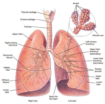
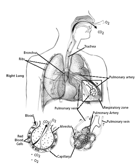
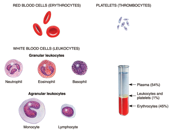
Red blood cells must be brought as closely as possible to the air we breathe, so that hemoglobin in the cells can give up waste carbon dioxide from cellular metabolism and take on oxygen. To accomplish this, the lungs have millions of tiny air sacs (alveoli) with very thin walls (alveolar membranes) containing microscopic blood vessels (capillaries) (see Figure 1 below). This anatomy of the lungs allows exposure of a large surface area of the blood to the air. Oxygen (O2) and carbon dioxide (CO2) gases diffuse (move) across the one cell thick alveolar membrane in opposite directions with the oxygen entering the blood and the carbon dioxide leaving it. This process is known as gas exchange.

Bronchi and lungs

Figure 1: Bronchi and lungs.

The gas exchange part of the lungs is known as the lung parenchyma (see Figure 2 below). Air is delivered to the parenchyma of the lungs through the bronchial tree — a repetitively branching tubular system for air conduction. It consists of the trachea, from which arises a right and left main (primary) bronchus to the right and left lungs. Smaller bronchi branch from the main bronchus of the right or left lung, then to smaller bronchi to the various lobes of the lungs, then to even smaller bronchi (bronchioles) that eventually reach the alveoli.

Mechanism of gas exchange

Figure 2: Mechanism of gas exchange.

How Respiration Is Impaired

Many diseases can affect breathing. The most useful classification of respiratory disorders is based on the manner in which the ability of air to come into contact with the hemoglobin in red blood cells (RBCs) is disrupted.

There are only two ways in which respiration can be impaired, regardless of the exact nature of the disorder:

1. Disease that prevents adequate amounts of air from reaching the gas-exchange level of the lungs (obstructive lung disease).

2. Disease of the lung tissue itself that reduces gas exchange (restrictive lung disease).

Thus, physicians have found it broadly useful to classify respiratory disorders as obstructive or restrictive, or a combination or the two.

Chronic Obstructive Lung Disease (COPD)

Chronic obstructive pulmonary disease (COPD) is the most common type of lung disease seen by the Social Security Administration. “Obstruction” refers to the fact that air flow in and out of the lungs is impeded. The three most frequent types of COPD in adults are:

1. Emphysema.

2. Chronic bronchitis.

3. Asthma.

In emphysema, lung tissue itself is destroyed. Damaged lung tissue forms non-functional spaces that trap air, and the lungs expand. The effect of these abnormalities is to obstruct air flow to and from the lungs. Emphysema and chronic bronchitis often occur together usually in people with a history of cigarette smoking.

Chronic bronchitis can also occur from long exposure to chemical fumes associated with a particular occupation. Inflammation of the inner surface of bronchi results from exposure to irritating substances. This inflammation, along with excessive secretion of bronchial mucous glands results in bronchial narrowing. Thus, bronchial tree resistance to air flow increases—there is obstruction of air flow.

Other less common causes of COPD include cystic fibrosis and bronchopulmonary dysplasia (BPD). These disorders, as well as asthma, have separate specific listings. See Can I Get Social Security Disability Benefits for Asthma? and Can I Get Social Security Benefits for Cystic Fibrosis?

Restrictive Lung Disease

The hallmark of restrictive lung disease is loss of usable lung volume, either due to:

1. Disease of the gas exchange part of the lungs (lung parenchyma), or

2. Some disorder outside of the lungs (extrapulmonary) that prevents air from adequately ventilating normal lung parenchyma.

Examples of Parenchymal Restrictive Lung Diseases

    • Infections (bacterial, fungal, viral, parasitic). Chronic infections that damage lung tissue, such as severe bronchiectasis or advanced pulmonary TB, could result in some degree of restrictive impairment.
    • Radiation, such as used for treatment of cancer, can damage lungs. Radiation lung damage is less of a problem than in the past, because modern equipment can direct therapeutic radiation in beams precisely delivered to the tumor. To the extent that this is impossible because of the size or location of the tumor, it is possible to have fibrotic lung damage caused by radiation.
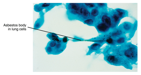
    • Inhalation of damaging substances into the lung. Pneumoconiosis is a non-specific term that refers to lung damage from inhaling small particles of some kind. Examples of pneumoconiosis include that caused by asbestos (asbestosis) (see Figure 3 below), coal dust (anthracosis), beryllium (berylliosis) silicon dust (silicosis), aluminum, iron, tin (stannosis) and talc. Inhalation of toxic chemicals—such as acidic fumes—can also damage lung tissue.

Asbestos particles in lung cells

Figure 3: Asbestos in lung cells.

  • Drug side-effects.
  • Autoimmune diseases. Autoimmune disorders such as sarcoidosis, scleroderma, systemic lupus erythematosus (SLE), rheumatoid arthritis (RA), polymyositis, ankylosing spondylitis, and mixed connective tissue disorders can cause pulmonary disease. In all of these disorders, some kind of immune system dysfunction damages the lungs. See Can I Get Social Security Disability Benefits for Lupus? and Can I Get Social Security Disability Benefits for Arthritis and Joint Damage?
  • Idiopathic diffuse interstitial pulmonary fibrosis (cryptogenic fibrosing alveolitis). This condition causes progressive scarring of the lungs on a microscopic level. This damage can result in decreased gas exchange capacity of the lungs, increasingly severe hypoxemia (lack of oxygen in the blood) and eventually death from respiratory failure. The rate of progression is highly variable, but median survival is less than 3 years. Some cases remain slowly progressive for a number of years, then are triggered by some unknown event to become rapidly more severe.
  • Other diseases. Examples of other diseases that can cause pulmonary fibrosis include alveolar proteinosis, acquired immunodeficiency syndrome (AIDS), acute respiratory distress syndrome (ARDS), amyloidosis, bone marrow transplantation, cancer, eosinophilic granuloma, eosinophilic pneumonia, lipoid pneumonia, genetic metabolic diseases caused by enzyme deficiencies (Gaucher’s disease, Niemann-Pick disease), Hermansky-Pudlak syndrome, pulmonary vasculitis, tuberous sclerosis, and neurofibromatosis. See Can I Get Social Security Disability Benefits for HIV / AIDS?

Examples of Extrapulmonary Restrictive Lung Diseases

    • Abnormal spinal curvatures. Abnormal curvatures of the spine can interfere with normal breathing movements. Scoliosis is a common spinal disorder, but will not compromise breathing until the major abnormal curve reaches about 60 degrees. Kyphosis is an abnormal curvature of the upper (thoracic) spine that causes a bent-forward or “hunched-over” posture. Kyphoscoliosis is a combination of both kyphosis and scoliosis.
    • Surgical resection. Removal of lung tissue limits the surface area available for gas exchange.
    • Spondyloarthropathies. Inflammatory disorders of the spine, such as ankylosing spondylitis, can make the spine more rigid by increased calcification of the spine and associated soft tissue ligaments. The ribs attach to the spine and their movement is impeded during expansion and relaxation of the chest during breathing. This limitation will decrease breathing capacity. See also Can I Get Social Security Disability Benefits for Back Pain?
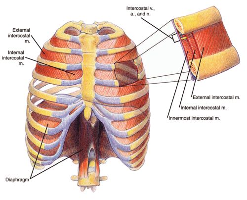
    • Thoracoplasty. Thoracoplasty, usually involving removal of one or more ribs, distorts the normal shape of the chest. The intercostal muscles between the ribs help expand the chest during inspiration. However, mechanical distortion of the chest wall is probably more important in decreasing ability to ventilate the lung during respiratory movements of the chest.
    • Obesity. Marked obesity can result in a significant reduction in ventilation capacity. Because of the weight of fat on the chest wall, the work of breathing is increased during inspiration. Furthermore, abdominal subcutaneous fat as well as intra-abdominal fat resists movement of the diaphragm—a respiratory muscle consisting of two sheets of muscle separating the chest and abdominal cavities (see Figure 4 below). The diaphragm must be able to move downward with inhalation in order to maximally expand the chest.

Chest cavity and diaphragm

Figure 4: Chest cavity and diaphragm.

  • Other important causes of restrictive respiratory impairment. Many disorders can result in weakness affecting the muscles of respiration (diaphragmatic muscles or intercostal muscles). Myasthenia gravis is an autoimmune disease that causes weakness through a biochemical interruption of the body’s ability to excite muscles, including the muscles of respiration. In fact, respiratory failure is usually the cause of death in myasthenia. There are a large number of muscle diseases (myopathies) that can affect respiration, although fairly rare in the general population.

Strokes and Breathing Problems

Strokes (cerebrovascular accidents, CVAs) are frequently adjudicated by the Social Security Administration. See Can I Get Social Security Disability Benefits After a Stroke? However, most of the time the treating source—even if a neurologist—does not consider the possibility of a breathing deficit resulting from the stroke. However, paralysis of half of the diaphragm is a definite possibility with resultant decreased ability to ventilate the lungs. Because treating sources, or consulting neurologists paid by the Social Security Administration, usually do not specifically address the possibility of diaphragmatic paralysis, SSA adjudicators are also likely to not even think of the possibility. Claimants themselves, following a stroke, may have thinking difficulties and neurological problems that occupy their attention as well as the attention of their family. The claimant cannot be counted on to mention breathing difficulty—especially since he or she may not even have symptoms in a resting state. The Social Security Administration adjudicator should ask to treating doctors, especially neurologists, regarding possible breathing deficits in post-stroke patients.

Pulmonary Function Studies

Pulmonary function study (PFS) is a general term that applies to any type of respiratory testing. The basic types of PFS are:

  • Spirometry
  • Arterial Blood Gas Study (ABGS)
  • Carbon Monoxide Diffusing Capacity (DLCO)

Shortness of breath is one of the most common allegations by claimants seeking Social Security disability benefits. The Social Security Administration pays for many PFS tests (especially spirometry) to address allegations of shortness of breath.

The results of these tests determine whether you qualify for disability benefits under a listing. See Winning Social Security Disability Benefits for Lung Disease by Meeting a Listing.

Spirometry

No pulmonary function study is more useful or more frequently performed than spirometry. Spirometry is the most important test for evaluating the severity of obstructive pulmonary disease. Spirometry requires you to inhale then exhale into a device called a spirometer. The device measures the volume of air that you can inhale and exhale and displays the result as a breathing curve on a graph called a spirogram.

If you have lung disease, but have not had a spirometry test, the Social Security Administration may arrange for you to have one. Even if you have had the test, the Social Security Administration may require you to be retested because the test must be administered in accordance with strict rules. Accurate testing must be done to assure you are treated fairly.

The test results must include the actual breathing curves. Spirometry test results in medical records often do not include the actual breathing curves, just the numerical results.

If there is no clinical evidence that you have significant lung disease and the reported spirometric values in your medical records are not significantly abnormal, the Social Security Administration will not send you for retesting. In fact, the Social Security Administration is not obligated to obtain spirometry on claimants who have no evidence in their medical records of a respiratory disorder, even if there are no spirometric values of any kind in the file.

Arterial Blood Gas Study (ABGS)

ABGS is the most important test used for the evaluation of the restrictive pulmonary disorders that involve a decrease in gas exchange—the parenchymal restrictive lung diseases. ABGS is performed on a sample of blood from the radial artery in the wrist or brachial artery in the arm, unlike most blood samples that are taken from a superficial vein just under the skin.

An ABG test checks how well your lungs are doing in moving oxygen into the blood and removing carbon dioxide from it. ABGS measures arterial oxygen pressure (PaO2), also known as oxygen tension, carbon dioxide pressure (PaCO2), and acidity (pH).

The Social Security Administration should not send you for ABGS, unless absolutely necessary to determine whether you are disabled, because it is invasive. Although complications are unusual, the person sticking a needle in the radial artery could damage the artery or other structures in the wrist; use of the brachial artery carries additional risk and should be done only when the radial artery cannot be used. The needle stick is also painful in either method.

Carbon Monoxide Diffusing Capacity (DLCO)

DLCO is a test used to evaluate of the severity of parenchymal restrictive lung diseases. It is of little value in assessing severity in COPD. The test measures how well your lungs can transfer carbon monoxide (CO) into the blood. By measuring how easily CO moves across the alveolar membrane of the lungs, doctors can deduce whether there is also a problem limiting the exchange of oxygen and carbon dioxide between blood and the atmosphere.

Two different methods are used for this test. The single-breath method is the one required by the Social Security Administration. This method requires you to take a breath of air containing a very small amount of carbon monoxide from a container while measurements are taken. (The other method, the steady-state method, requires you to breathe air containing a very small amount of carbon monoxide. The amount of carbon monoxide in the breath you exhale is then measured.)

DLCO can be abnormal in emphysema, if the disease is very advanced, because lung tissue is damaged. However, DLCO is not an accurate enough means of determining the severity of emphysema; spirometry is more appropriate. In some instances, more applicable to clinical medicine than disability determination, DLCO testing can help distinguish between emphysema and asthma, because asthma will have normal values.

Continue to Winning Social Security Disability Benefits for Lung Disease (Chronic Pulmonary Insufficiency) by Meeting a Listing.

Schizophrenia

By Applying for disability benefits when you have:

Can I Get Social Security Disability Benefits for Schizophrenia?

  • How Does the Social Security Administration Decide if I Qualify for Disability Benefits for Schizophrenia?
  • About Schizophrenia and Disability
  • Winning Social Security Disability Benefits for Schizophrenia by Meeting a Listing
  • Residual Functional Capacity Assessment for Schizophrenia
  • Getting Your Doctor’s Medical Opinion About What You Can Still Do

How Does the Social Security Administration Decide if I Qualify for Disability Benefits for Schizophrenia?

If you have schizophrenia, Social Security disability benefits may be available. To determine whether you are disabled by schizophrenia, the Social Security Administration first considers whether the schizophrenia is severe enough to meet or equal a listing at Step 3 of the Sequential Evaluation Process. See Winning Social Security Disability Benefits for Schizophrenia by Meeting a Listing. If your schizophrenia is not severe enough to equal or meet a listing, the Social Security Administration must assess your residual functional capacity (RFC) (the work you can still do, despite schizophrenia), to determine whether you qualify for disability benefits at Step 4 and Step 5 of the Sequential Evaluation Process. See Residual Functional Capacity Assessment for Schizophrenia.

About Schizophrenia and Disability

What Is Schizophrenia?

Schizophrenia is a serious mental disorder for which there is no cure. Schizophrenia affects about 1% of the adult population. There are various types of schizophrenia, i.e., disorganized, catatonic, residual, undifferentiated, and paranoid. Any of these forms of schizophrenia may entitle a person to disability benefits.

Schizophrenia appears before age 45, usually in the adolescent or young adult. The person exhibits a definite deterioration from a previous level of functioning in social, work, and personal life. Psychological abnormalities are diverse, affecting:

  • Thought.
  • Perception.
  • Emotion (affect).
  • Identity.
  • Ability to plan and carry out purposeful activity.
  • Cognition (ability to think, learn, and understand).
  • Sociability. The person may experience a tendency to withdraw from the outside world and a preoccupation with illogical ideas, fantasies, or delusions.
  • Movement. The person may exhibit a decrease, increase, or bizarreness of movements or postures.

The prevailing view of schizophrenia has been that the psychotic features are the most important and that cognitive deficits are secondary. However, the latest research suggests that the cognitive deficits, which arise from various brain abnormalities, are the more fundamental cause of mental abnormalities in schizophrenia. In fact, there is reason to think that subtle brain abnormalities appear before a child is born and gradually increase in severity.

Causes of Schizophrenia

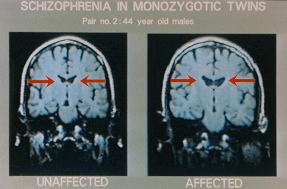
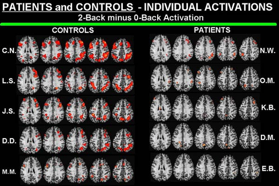
The disorder involves biochemical dysfunction in the brain, but no one can as yet describe the mechanisms involved. Numerous abnormalities are found in the brains of those with schizophrenia (see Figures 2 and 4 below).

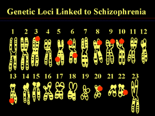
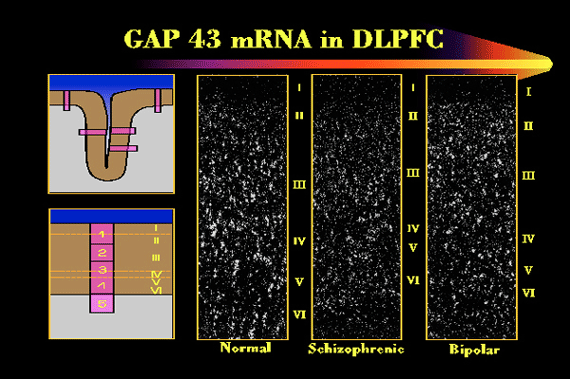
There is also a complex genetic predisposition that no one is close to understanding. It is thought that about 40% of cases of schizophrenia are inherited, and 60% occur spontaneously. Recent research suggests that schizophrenia may result from a large number of genetic abnormalities (see Figures 1 and 3 below). Consequently, there is no chance of a single cause and cure for schizophrenia.

Linkage Studies and Schizophrenia

Figure 1: A so-called “linkage study,” showing a number of places in the human genome where pieces of DNA are inherited along with risk for the illness. It shows one of each of the 23 pairs of chromosomes, and the red dots indicate regions where a piece of DNA has been shown to be inherited along with the risk for schizophrenia in certain families and certain studies.

MRI Scans and Schizophrenia

Figure 2: MRI scans of identical twins. The twin on the right has schizophrenia; the twin on the left is healthy. Even to the unprofessional eye, there are obvious differences, a systematic and consistent variation between the affected and the unaffected twin in the gross anatomy of the brain. Red arrows point to enlarged ventricles in the affected twin.

Schizophrenia and mRNA

Figure 3: Each white dot represents cells in a particular part of the brain. A patient with schizophrenia is compared to an individual with another psychiatric illness, bipolar disorder, and to a normal subject. The white dots show the turning-on of a gene that is the blueprint for a protein related to the process by which cells adapt themselves to a changing environment. I–VI represent layers of the cerebral cortex.

Schizophrenia PET Scans

Figure 4: PET scans of five normal individuals (left); each row is one person, and each image is a slice from five different levels of the person’s brain. The red areas show regions of the brain that are activated when a person performs a memory task. In PET scans of five individuals with schizophrenia (right), each row represents a different person, with comparable slices. Clearly, the patients with schizophrenia do not generate the dramatic brain activity in the circuits of the brain critical to the memory task.

Phases of Schizophrenia

Schizophrenia usually has 3 phases.

  • First, there is the prodromal phase. The duration of this phase is variable. The person shows a clear deterioration from the previous level of functioning in multiple areas of life—mental, occupational, personal, and social. The “change in personality” is often noticed by friends and relatives, as the person withdraws from usual activities, engages in less effective and increasingly strange behavior and experiences difficulty with clear and logical communication with others.
  • Next is the active or progressive phase. In this phase, overt psychotic symptoms become obvious. The impairments present in the prodromal phase become more severe. The individual increasingly withdraws. Strange behavior may become bizarre or completely disorganized, and odd perceptions and thought develop into hallucinations and delusions. Abnormalities in the form and content of thought produce further confusion and difficulty communicating with others. Transition from the prodromal to the active phase is often triggered by stress that the person cannot cope with. For example, such a stressor might be a serious physical illness or change in an important relationship with another person. Or it might be something that a normal person would consider trivial.
  • A residual phase follows the active phase. Psychotic symptoms persist to some degree, but the accompanying emotions are not as intense. (The residual phase should not be confused with the residual type of schizophrenia, so-called because an active, florid phase of the illness is missing.) Unfortunately, normal emotional responsiveness tends to be subdued also. Residual functional impairment may remain severe.

The majority of claimants seeking disability benefits for schizophrenia are in the residual phase, and in some degree of remission on neuroleptic drugs which must be taken indefinitely. However, schizophrenia almost never goes into complete remission.

Cognitive Impairment With Schizophrenia

It is very important for the Social Security Administration to pay attention to cognitive dysfunction when assessing whether a person with schizophrenia is disabled. The Social Security Administration and many psychiatrists and psychologists still emphasize the psychotic features (hallucination, delusions) of the disease. So if a claimant’s psychotic features are controlled by medication, the Social Security Administration is not likely to test memory or other cognitive functions. Fewer than 10% of patients with schizophrenia ever get a regular job or live independently. At least 90% of people with schizophrenia are apparently incapable of working on any sustained basis.

Side Effects of Schizophrenia Medications

The drugs taken to lessen the symptoms of schizophrenia can cause side effects. The severity varies from person to person. In the early phase of drug therapy, symptoms may include muscle spasms, tremors, dry mouth, drowsiness, blurry vision, and restlessness. Changing the type of medication or the dosage can help control these symptoms.

Older medications were associated with a risk of the patient developing tardive dyskinesia—a disorder characterized by involuntary movements, especially of the lips, tongue, and mouth. Newer medications, such as clozapine (Clozaril), olanzapine (Zyprexa), quetiapine (Seroquel) and resperidone (Resperdal) are associated with other risks. For example, clozapine can cause a decrease in white blood cells and greatly weaken the body’s defense against dangerous infections. Clozapine can also cause seizures, inflammation of the heart (myocarditis), low blood pressure (hypotension), and other side effects. Olanzapine (Zyprexa) can cause weight gain and Parkinsonian movement disorders. Anyone taking antipsychotic medications must be monitored by a doctor.

If the claimant is having medication side-effects, a Social Security Administration psychiatrist or other medical doctor, rather than a Social Security Administration psychologist, should evaluate at least that part of the claim.

Information from Family and Friends Is Crucial to Disability Determination

Claimants with chronic schizophrenia living with family members are most likely to be improperly denied Social Security disability benefits. It is critically important for family members or other caregivers to provide the Social Security Administration with as detailed information as possible about specific tasks the claimant can or cannot do.

Mental health clinics will often refuse to provide the Social Security Administration with clinical records that are useful in evaluating how a mental disorder has developed over time. They might simply write a letter summarizing what they think they Social Security Administration needs to know. In some instances, the Social Security Administration is forced to fall back on purchasing a consultative mental status examination in which the examining psychiatrist or psychologist has limited time to determine the details of daily functional capacity. The Social Security Administration should ask the treating psychiatrist (or psychologist) about work-related abilities for at least unskilled work and how these conclusions match with the corresponding limiting mental symptom.

If the claimant is receiving medication, information about side-effects must come from a medical doctor, because a psychologist is not competent to evaluate that matter. However, it is also important for the same kind of information and opinions to be obtained from family or other caregivers, to make sure that nothing is missed. The caregivers live with the claimant; they may have noticed important facts that can be brought to the treating psychiatrist’s attention and to the Social Security Administration.

Continue to Winning Social Security Disability Benefits for Schizophrenia by Meeting a Listing.

Hepatitis C & liver disease

By Applying for disability benefits when you have:

Can I Get Social Security Disability Benefits for Hepatitis or Other Chronic Liver Diseases?

  • How Does the Social Security Administration Decide if I Qualify for Disability Benefits for Chronic Liver Disease?
  • About Chronic Liver Disease and Disability
  • Winning Social Security Disability Benefits for Liver Disease by Meeting a Listing
  • Residual Functional Capacity Assessment for Chronic Liver Disease
  • Getting Your Doctor’s Medical Opinion About What You Can Still Do

How Does the Social Security Administration Decide if I Qualify for Disability Benefits for Chronic Liver Disease?

If you have any type of chronic liver disease, including chronic viral hepatitis, Social Security disability benefits may be available. To determine whether you are disabled by your chronic liver disease, the Social Security Administration first considers whether your liver disease is severe enough to meet or equal a listing at Step 3 of the Sequential Evaluation Process. See Winning Social Security Disability Benefits for Liver Disease by Meeting a Listing. If you meet or equal a listing because of chronic liver disease, you are considered disabled. If your liver disease is not severe enough to equal or meet a listing, the Social Security Administration must assess your residual functional capacity (RFC) (the work you can still do, despite your liver disease), to determine whether you qualify for benefits at Step 4 and Step 5 of the Sequential Evaluation Process. See Residual Functional Capacity Assessment for Liver Disease.

About Chronic Liver Disease and Disability

What Is Chronic Liver Disease?

Chronic liver disease is characterized by liver cell necrosis (death), inflammation, or scarring (fibrosis or cirrhosis) due to any cause that persists for more than 6 months.

Examples of Chronic Liver Disease

Examples of chronic liver disease include, but are not limited to:

  • Chronic hepatitis;
  • Alcoholic liver disease;
  • Non-alcoholic steatohepatitis (NASH);
  • Primary biliary cirrhosis (PBC);
  • Primary sclerosing cholangitis (PSC);
  • Autoimmune hepatitis;
  • Hemochromatosis (liver damage from iron overload, resulting from a defect in the body’s ability to metabolize iron properly);
  • Drug-induced liver disease;
  • Wilson’s disease(a rare genetic disease caused by a defect in the body’s ability to metabolize copper); and
  • Serum alpha-1 antitrypsin deficiency.

There are many other possible causes of liver disease, such as metabolic disorders (e.g., cystic fibrosis, porphyria, glycogen storage disease), vascular diseases (e.g., polymyalgia rheumatica, polyarteritis nodosa), parasitic infections (particularly roundworms, tapeworms, and flatworms), bacterial and fungal infections, toxins and drugs, and autoimmune hepatitis.

Some cases of liver failure are caused by an unknown agent.

Signs and Symptoms of Chronic Liver Disease

Signs of chronic liver disease may include:

  • Jaundice (yellowing of the skin),
  • Enlargement of the liver and spleen,
  • Ascites (fluid in the abdomen),
  • Peripheral edema (swelling of extremities), and
  • Altered mental status.

Symptoms may include:

  • Pruritis (itching),
  • Fatigue,
  • Nausea,
  • Loss of appetite, or
  • Sleep disturbances.

Symptoms of chronic liver disease may have a poor correlation with the severity of liver disease and functional ability.

Laboratory Findings Associated With Liver Disease

With liver disease, blood tests may show increased liver enzymes, increased serum total bilirubin, increased ammonia levels, decreased serum albumin, and abnormal coagulation studies, such as increased International Normalized Ratio (INR) or decreased platelet counts.

Abnormally low serum albumin or elevated INR levels indicate loss of liver function, with increased likelihood of cirrhosis and associated complications. However, other abnormal lab tests, such as liver enzymes, serum total bilirubin, or ammonia levels, may have a poor correlation with the severity of liver disease and functional ability.

A liver biopsy may demonstrate the degree of liver cell necrosis, inflammation, fibrosis, and cirrhosis. If you have had a liver biopsy, the Social Security Administration will make every reasonable effort to obtain the results; however, it will not pay for you to have one.

Imaging studies (CAT scan, ultrasound, MRI) may show the size and consistency (fatty liver, scarring) of the liver and document ascites (fluid in the abdomen).

Chronic Viral Hepatitis Infections

Hepatitis means inflammation of the liver. If lasting more than about 6 months, it may be termed chronic. Prolonged hepatitis can lead to cirrhosis.

Although there are several different types of viral hepatitis infections, chronic viral hepatitis infections are commonly caused by hepatitis C virus (HCV), and to a lesser extent, hepatitis B virus (HBV). Usually, these are slowly progressive disorders that persist over many years during which the symptoms and signs are typically nonspecific, intermittent, and mild (for example, fatigue, difficulty with concentration, or right upper quadrant pain).

The spectrum of these chronic viral hepatitis infections ranges widely. Some patients may have no symptoms; others may have only mild to moderate symptoms. Some may develop cirrhosis, end stage liver disease with the need for liver transplantation, or liver cancer.

Chronic Hepatitis B (HBV) Infection

Chronic HBV infection is diagnosed by the detection of hepatitis B surface antigen (HBsAg) in the blood for at least 6 months. The goal of treatment is to suppress HBV replication and thereby prevent progression to cirrhosis and end stage liver disease. Treatment usually includes a combination of interferon injections and oral antiviral agents.

The hepatitis B virus is a highly infectious organism. Worldwide, there are hundreds of millions of carriers. Millions of individuals die yearly from hepatitis B viral infections. HBV may induce a form of liver cancer known as hepatocellular carcinoma that kills many hundreds of thousands of people worldwide every year.

In the U.S., probably less than 1% of the population are carriers. This is much lower than in some other parts of the world where the majority of individuals have been infected. High-risk groups for infection are male homosexuals, IV drug users, and individuals working in healthcare who are exposed to blood or blood products.

There is about a 10% chance of infection of a person who suffers an accidental stick with a needle contaminated with the blood of a HBV carrier. Donated blood is routinely screened for hepatitis B to prevent transmission by transfusion, but this doesn’t protect health workers exposed to blood in the course of a patient’s care. In addition to blood, a carrier has virus in semen, saliva, vaginal secretions, sweat, tears, urine, and breast milk.

If swallowed, the virus is destroyed by pancreatic enzymes in the digestive tract which probably explains why oral transmission is rare and transmission in food has not been documented. In the rare cases of oral transmission, infection probably occurred because of breaks in the oral mucosa or gingiva that allowed the virus to directly enter the bloodstream.

Transmission of the virus from blood or other bodily fluids through small cuts or abrasions in the skin or mucous membranes of which a person is unaware may explain why as many as 50% of infected individuals have no history of high-risk exposure. As many as 80% to 90% of infected individuals are unaware they are carriers.

Maximum symptoms occur at about 3 to 5 months after initial infection and correspond to the greatest liver inflammation as reflected by a peak in transaminase liver enzymes. Once infected, some individuals may die within a matter of weeks from severe and aggressive hepatitis and liver failure, but this outcome involves less than 1% of cases. Most patients (90% to 95%) will have a full recovery, and 5% to 10% will become chronic carriers of the virus.

Carriers with no symptoms who also have normal levels of transaminase enzymes have a good prognosis, because they have no continuing hepatitis. Those who have abnormal transaminases indicative of continuing hepatitis have a poorer prognosis even if they have no symptoms. Such carriers are more likely to develop chronic persistent hepatitis (CPH), chronic active hepatitis (CAH), and hepatocellular carcinoma.

Chronic Hepatitis C (HCV) Infection

Chronic HCV infection is diagnosed by the detection of hepatitis C viral RNA in the blood for at least 6 months. Hepatitis C can be transmitted in blood and can cause both acute and chronic liver inflammation, as well as cirrhosis of the liver. Along with alcohol abuse, hepatitis C is the most common cause of cirrhosis. At least 4 million people in the U.S. are infected, and many are unaware that they are infectious. Risk of infection is increased by sexual intercourse with infected individuals and by intravenous drug use. About half of infected individuals progress to chronic active hepatitis (CAH) and have an associated increased risk of liver cancer.

Treatment usually includes a combination of interferon injections and oral ribavirin. Whether a therapeutic response has occurred is usually assessed after 12 weeks of treatment by checking the HCV viral load. A substantial reduction in HCV viral load (also known as early viral response, or EVR) predicts a sustained viral response with completion of treatment. Combined therapy is commonly discontinued after 12 weeks when there is no early viral response, since in that circumstance there is little chance of obtaining a sustained viral response (SVR). Otherwise, treatment is usually continued for a total of 48 weeks.

Combined interferon and ribavirin treatment may have significant adverse effects that may require dosing reduction, planned interruption of treatment, or discontinuation of treatment. Adverse effects may include: anemia (ribavirin-induced hemolysis), neutropenia (low neutrophil [type of white blood cell] count), thrombocytopenia (low platelet count), fever, cough, fatigue, myalgia (muscle pain), arthralgia (joint pain), nausea, loss of appetite, pruritis (itching), and insomnia. Behavioral side effects may also occur. Influenza-like symptoms are generally worse in the first 4 to 6 hours after each interferon injection and during the first weeks of treatment. Adverse effects generally end within a few days after treatment is discontinued.

Chronic Active Hepatitis

Chronic active (aggressive) hepatitis (CAH) is a very serious disorder whose specific cause is unknown, but may involve some abnormal immune response by the liver to a particular agent. If the CAH is related to intake of a drug, it may rapidly improve with withdrawal of that drug. However, cases associated with viral infections have a much poorer prognosis. Overall, CAH has a high mortality despite treatment, and sometimes can be associated with problems in organ systems other than the liver. These include arthralgia (joint pains), anemia, fibrosis of the lungs, intestinal inflammation, and kidney disease.

Cirrhosis

The most frequent kind seen by the Social Security Administration is cirrhosis of the liver caused by the abuse of alcohol. The term cirrhosis refers to present or past evidence of hepatic (liver) cell necrosis (death), fibrosis (replacement of normal cells with fibrous tissue), and areas of regenerating nodules of liver tissue attempting to recover from previous damage.

Tissue for microscopic evaluation must be removed from the liver (biopsy), which is the only way a certain diagnosis of cirrhosis can be made. The Social Security Administration does not purchase liver biopsies on claimants. When they are done by a treating source, they are usually percutaneous—a needle is inserted into the liver through the skin between the right lower ribs under local anesthesia and can be done at the bedside.

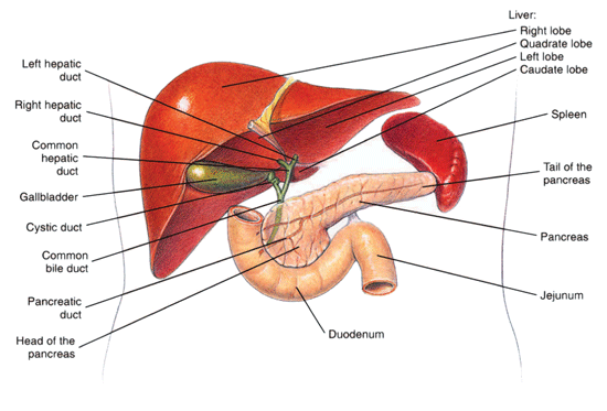
The liver makes and secretes bile into the bile ducts, and these ducts in turn carry bile to the gallbladder for storage, and also to the small intestine (see Figure 1 below). Bile assists in the absorption of fats from the intestine. Biliary cirrhosis results when normal bile flow is interrupted and the liver cells are exposed to it. Primary biliary cirrhosis is usually found in older females and involves obstruction to bile flow in the small biliary ducts inside the liver itself. Secondary biliary cirrhosis involves obstruction of the larger ducts outside the liver (extrahepatic ducts) with a consequent back-pressure of bile into the liver. Gallstones may produce an obstruction, or stricture related to previous surgery, or rarely an obstructing cancer.

Liver, pancreas, and duodenum

Figure 1: The liver, pancreas, and duodenum.

Toxins as a Cause of Hepatitis

Many toxic substances can cause hepatitis, the most prominent of which is excessive alcohol intake. People vary greatly in the efficiency with which their liver can metabolize alcohol, and there is no way to predict individual capacity in this regard. By far, alcoholic hepatitis is the most frequent form of hepatitis and is usually acutely superimposed upon a more chronic underlying cirrhosis. As a patient is detoxified in a treatment facility, the alcoholic hepatitis improves with progression of residual chronic cirrhosis. Some cases of acute alcoholic hepatitis are fatal and cirrhosis eventually results in the death in many others.

Acute Hepatitis

Alcoholics, in particular those who have been binge drinking, can have an episode of acute alcoholic hepatitis superimposed on an underlying chronic liver disease. When hospitalized where alcohol is unavailable and treated, the acute hepatitis can improve. In these claims, the Social Security Administration can be expected to hold the claim for a few months to determine what degree of improvement will occur with treatment of the acute episode.

Claimants with acute viral hepatitis will be treated similarly, because most will improve over the short term.

Continue to Winning Social Security Disability Benefits for Liver Disease by Meeting a Listing.

Ischemic heart disease

By Applying for disability benefits when you have:

Can I Get Social Security Disability Benefits for Ischemic Heart Disease?

  • How Does the Social Security Administration Decide if I Qualify for Disability Benefits for Ischemic Heart Disease?
  • About Ischemic Heart Disease and Disability
  • Winning Social Security Disability Benefits for Ischemic Heart Disease by Meeting a Listing
  • Residual Functional Capacity Assessment for Ischemic Heart Disease
  • Getting Your Doctor’s Medical Opinion About What You Can Still Do

How Does the Social Security Administration Decide if I Qualify for Disability Benefits for Ischemic Heart Disease?

If you have ischemic heart disease, Social Security disability benefits may be available to you. To determine whether you are disabled by your ischemic heart disease, the Social Security Administration considers whether your heart disease is severe enough to meet or equal a listing at Step 3 of the Sequential Evaluation Process. See Winning Social Security Disability Benefits for Ischemic Heart Disease by Meeting a Listing. If you meet or equal a listing because of ischemic heart disease, you are considered disabled.

If your ischemic heart disease is not severe enough to equal or meet a listing, the Social Security Administration must assess your residual functional capacity (RFC) (the work you can still do, despite your heart disease), to determine whether you qualify for disability benefits at Step 4 and Step 5 of the Sequential Evaluation Process. See Residual Functional Capacity Assessment for Ischemic Heart Disease.

About Ischemic Heart Disease and Disability

What Is Ischemic Heart Disease?

Ischemic heart disease (IHD) is a major medical problem disabling and killing millions of people in the U.S. yearly, and therefore a frequent basis for allegations of disability. Heredity plays an important role, but so does lifestyle. Many IHD deaths are preventable and associated with poor control of hypertension and diabetes mellitus, smoking, lack of exercise, obesity, and poor diet resulting in elevated blood lipids (fats such as cholesterol).

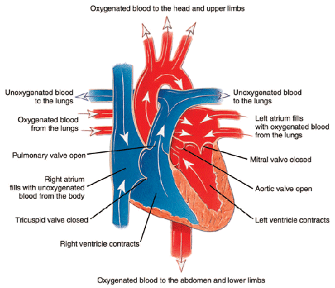
Ischemia means that the heart (or other body tissue) is not receiving enough oxygen to function normally. The oxygen demands of the heart change with heart rate (see Figure 1 below). So increased heart rates require increased blood flow to the heart to deliver the oxygen and glucose needed for the extra work. Ischemia not present at rest may appear when the heart rate increases, and this is the basis for cardiac stress testing. See Cardiac Stress Testing.

Heart Contraction

Figure 1: The path of oxygenated and unoxygenated blood in the heart during contraction.

Causes of Ischemic Heart Disease

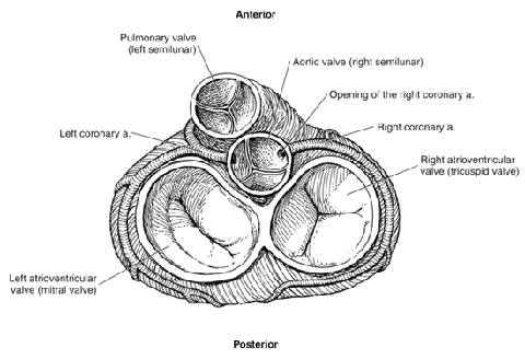
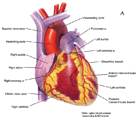
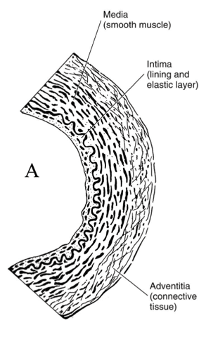
Most IHD results from obstruction of the coronary arteries that supply the myocardium (heart muscle) with blood. This condition is known as coronary artery disease (CAD). Other cardiac abnormalities can also cause myocardial ischemia. The underlying mechanism is still the same: restriction of blood flow to the myocardium. For example, marked narrowing of the aortic valve (aortic stenosis) (see Figure 2 below) can cause angina (chest pain) because the coronary arteries originate right above the valve (see Figure 3 below) and may not receive sufficient blood flow to supply the heart. However, the great majority of claimants have myocardial ischemia because of CAD.

Closed heart valves

Figure 2: Closed heart valves.

Anterior view of the heart

Figure 3: Anterior view of the heart, including main arteries.

How Severe Does Coronary Artery Disease Have to Be to Produce Ischemia?

Blood flow to the myocardium served by a particular coronary artery is not significantly decreased until a lesion blocks about 70% of the diameter of the artery. It is frequently possible for a person to be comfortable at rest with a 90% blockage in a large epicardial artery.

Lesions of less than 50% are generally considered to not be significant CAD, because the heart will still be able to obtain sufficient blood flow both at rest and with exercise. However, this conclusion must be made in context of all of the medical evidence. Long segments of obstruction rather than discrete lesions, involvement of the left main coronary artery, or involvement of multiple arteries could cause significant ischemia with blockages as low as 50%.

Detecting Ischemia

Ischemia can be indicated by:

  • Electrocardiograms (ECGs, ECGs);
  • Imaging studies that show the movement of the cardiac ventricular walls;
  • Imaging studies that show relative blood flow within the heart muscle; and
  • Imaging studies that measure amounts of blood moved by the heart’s ventricles.

Cardiac Stress Testing

Cardiac stress testing raises heart rate to determine:

1) Whether ischemia can be induced,

2) The level of exertion producing ischemia, and

3) The severity and location of the ischemia.

Cardiac stress testing is also used to determine the exercise capacity of individuals in regard to physical conditioning, but the Social Security Administration never purchases testing for that purpose alone.

The only type of cardiac stress testing mentioned by the Social Security Administration is exercise stress testing, in which physical exertion is used to raise the patient’s heart rate.

In exercise stress testing, your baseline ECG is obtained. Then you begin with a low level of exercise, either by walking on a treadmill, or pedaling a stationary bicycle. Every two to three minutes, the level of exertion is increased. At each stage of exercise, your pulse, blood pressure and ECG are monitored are recorded, along with any symptoms you may be experiencing.

The exertion level is gradually increased every 2 to 3 minutes until you cannot keep up any longer, or until your symptoms (chest pain, shortness of breath, or lightheadedness) prevent further exercise, or until changes on your ECG indicate a cardiac problem.

There are other means of raising heart rate for testing purposes, such as pharmacologic stress testing in which drugs are used to raise the heart rate. Pharmacologic stress testing can be used with patients who, for some reason, cannot perform exercise stress testing.

Both exercise testing and pharmacologic testing can be used in conjunction with various types of imaging studies of the heart’s response to increased heart rate—such as radionuclide scans or echocardiograms that are done at the same time.

Left Ventricular Ejection Fraction

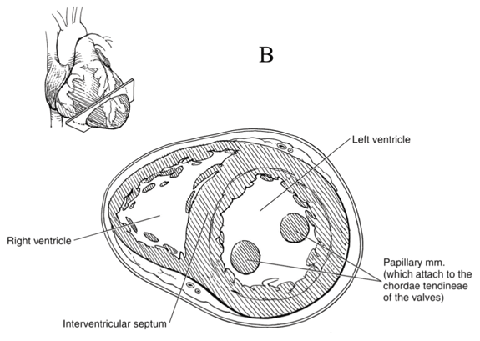
The most important performance test for blood moved by cardiac ventricles (see Figure 4 below) is the percentage of blood pumped out of the left ventricle (LV) with each beat—the left ventricular ejection fraction (LVEF). The left ventricle is of major interest, because it pumps blood into the systemic circulation that supplies the tissues of the body and is the major site of damage by ischemic heart disease, such as heart attacks.

Cross-section of the ventricles of the heart

Figure 4: Cross-section of the ventricles of the heart, which are important to blood movement.

The LVEF is normally about 55-65%, and is not usually considered abnormal unless it falls under 50%.

Angina Pectoris and Ischemic Heart Disease

What is Angina Pectoris?

When the myocardium receives inadequate blood flow, sensory nerve fibers (cardiac afferents) from the heart to the brain carry impulses for pain known as angina pectoris. The Social Security Administration also refers to angina as “chest discomfort of myocardial ischemic origin.”

In a significant number of people with myocardial ischemia, the pain activation system is defective for no obvious reason. These people have no warning when blood flow to the heart is inadequate and they are at particular risk for sudden death. Without warning pain, such individuals may continue to exercise after the onset of ischemia. Ischemic heart muscle is electrically unstable, so that it carries an increased risk of a lethal arrhythmia.

Angina and Disability

Angina is relevant to disability determination. To meet the listing for ischemic heart disease, you must have symptoms due to myocardial ischemia. See Winning Social Security Disability Benefits for Ischemic Heart Disease by Meeting a Listing. Angina is one such symptom. See Symptoms Due to Myocardial Ischemia.

Angina is also relevant to disability determination because chest pain limits exertion. In disability adjudication, the Social Security Administration frequently must evaluate the status of claimants after a myocardial infarction. See Heart Attacks (Myocardial Infarctions or MIs).

Many allege chest pain that may or may not be of cardiac origin.

However, there are too many unknown variables, conflicting descriptions, and other confounding factors to use the absence or presence of chest pain—even classic, angina-compatible chest pain—as controlling either treatment or disability decision-making.

Objective Data Are Necessary for Diagnosis of Angina

Objective data (test results) supporting the angina diagnosis are necessary. A diagnosis of angina based on a patient’s description alone is not reliable. Many claimants who describe chest pain that could be angina based on the description they provide (angina-compatible chest pain), turn out to have nothing wrong with their hearts after thorough examination.

A number of medical conditions can cause chest pain closely resembling angina. Esophageal spasm, for example, can feel just like angina—even be relieved with drugs used to treat angina—except for one thing: the absence of relation to exertion. A good history of chest pain that covers the possible non-cardiac types of pain requires a careful approach and professional medical training. Shortness of breath may occur with angina, but shortness of breath alone is not angina.

The Social Security Administration must obtain cardiac exercise testing on claimants with angina-compatible chest pain when there is no other means of allowance and there is no contraindication to such testing. See Cardiac Stress Testing.

Characteristics of Angina

Angina is described in terms of:

  • Location,
  • Quality,
  • Precipitating causes,
  • Duration, and
  • Mode of relief.

The general characteristics of these factors are as follows:

Location – Central Chest (Substernal)

Angina pain sometimes radiates down one or both arms or up into the neck or jaw. Angina is much less likely if the pain is only outside of the central chest area, such as in the jaw alone, or abdomen. Pain in a small area on the side of the chest is probably not angina. The more unusual the location of pain, the less likely that angina is actually the cause. However, if other characteristics fit the diagnosis of angina then it should be accepted despite an atypical location—especially if there is objective evidence of significant ischemic heart disease. In fact, the Social Security Administration is willing to accept atypical locations—even pain isolated to a hand, jaw, or neck—if everything else fits the angina diagnosis.

Quality – Dull, Aching, Squeezing, Heavy

Most claimants describe their pain as “sharp.” For some reason, this is the first word that enters the mind for many people when asked the quality of a pain. Therefore, a diagnosis of angina should not be discarded because a claimant uses the word “sharp.” Less-educated claimants are particularly likely to use words for the quality of chest pain that actually do not describe what they are experiencing. Because of the subjective nature of “quality”—what something feels like—the Social Security Administration adjudicator should only reject the characteristic if the quality described is something that is never associated with angina.

Rhythmic pain is never angina. Rhythmic pain is something that rapidly varies in intensity, especially stabbing, jabbing and poking sensations that quickly come and go (also see the discussion of Duration below). It is important for the interviewer to carefully communicate with the patient in this regard to make sure that there is mutual understanding of the meaning. For example, sticking or “pins-and-needles” sensations are also not compatible with angina.

Precipitating Causes – Exertion, Emotion

Exertion and emotion (excitement, anxiety, fear) raise heart rate, which can induce cardiac ischemia and the onset of ischemic chest pain—angina. Chest pain that always occurs with particular activities, such as walking a certain distance or climbing a certain number of stairs is characteristic of angina.

Chest pain with exertion does not always indicate angina. Chest pain that occurs randomly, sometimes during exertion and sometimes during rest, does not suggest ischemia since the cause-and-effect element is missing.

Chest pain associated with emotion, but occurring at rest, could be angina. On the other hand, chest pain that occurs only with emotion and not with exertion is suspect for being of non-cardiac origin.

Although there should be a general cause and effect relationship between angina and exertion or emotion, that does not mean that angina will always occur at a specific level of exertion since multiple factors (e.g., emotional state, physical health, drugs, temperature) can influence it. The absence of chest pain during exercise testing is not sufficient, in itself, to rule out ischemic heart disease. Nor is it sufficient to eliminate a claimant’s subjective report of chest pain during activities of daily living from consideration. Also, some individuals may show evidence of ischemia during stress testing, but have no pain.

Ischemia and reported symptoms are not predictably associated. Similarly, ischemia at any particular level of exercise cannot be closely predicted on the basis of the underlying coronary artery lesions.

The important piece of information needed for either treatment or disability determination is objective, exercise-induced ischemia.

Vasospastic Angina

There is an important exception to the relationship between exertion and ischemic cardiac chest pain: vasospastic angina (variant angina) occurs when a coronary artery narrows in a particular location, thereby decreasing blood flow to heart muscle past the point of narrowing. The word “vasospastic” means that the narrowing is temporary, and usually results from some irritative focus that causes the smooth muscle inside the coronary artery to constrict at a specific place. The irritative focus is something that triggers the muscle spasm and that is often the fibro-fatty, sometimes calcified, plaque of coronary artery disease.

Drugs like cocaine are particularly likely to induce coronary artery spasm. In fact, cocaine can cause such severe constriction that blood flow is completely cut off to an area of the heart, with a resultant heart attack in a young person who would otherwise not have such an event. The risk is probably even greater for a middle-aged person who already has an irritative focus like a fatty lesion in a coronary artery and then also takes cocaine.

Angina in the Resting State

It is possible to have cardiac ischemia so severe that angina occurs in the resting state. But these people are so ill that they have been evaluated for heart disease by their treating physicians and severe abnormalities have been documented. Angina occurring in the resting state either results in medical treatment or death. No one with angina at rest is going to be in that state very long without medical care. Furthermore, angina in such cases will still be characteristically precipitated by exertion and worsened by exertion, so that important cause-and-effect relationship is still intact.

Duration

Typical angina lasts only a couple of minutes after the precipitating cause is stopped. It is quite common for claimants to say that their chest pain lasts for hours at a time on a regular basis. This is not anginal chest pain. Chest pain lasting for hours is more compatible with a heart attack than angina. Even if a person has particularly severe and worsening angina—a condition known as unstable angina—there is no way such prolonged angina could be occurring on a regular basis, because the individual would be dead or in a hospital.

Attempting to continue exertion with angina will cause further worsening until the person stops the inducing activity, which will happen quickly after the onset of pain; no one ignores angina.

Chest pain lasting less than a minute is usually not angina. Chest pain lasting less than 10 seconds or more than 30 minutes would be considered non-anginal by most cardiologists. In the unusual cases in which 30 minute durations are caused by ischemic heart disease, they are more characteristic of unstable angina, than typical angina. Unstable angina is a serious condition and not something a person has very long without coming under medical care. Nevertheless, the Social Security Administration is usually generously flexible in this regard. If all of information is supportive of the diagnosis, durations of up to 30 minutes will generally be accepted in disability determination as angina, rather than requiring a more realistic 3 to 5 minutes of duration. Perhaps this is because the Social Security Administration realizes that many people are poor at estimating the time their chest pain lasts. Of course, the optimum situation is one in which patients have actually measured the duration of pain after cessation of the precipitating cause, but claimants usually have not done so.

Relief of Angina

Angina caused by exertion is relieved by rest; emotionally induced angina is relieved by lessened emotional arousal.

A number of long-acting drugs improve cardiac ischemia and decrease the over-all frequency and severity of anginal attacks. For example, beta-blocker drugs like propranolol will keep the exertional and emotional heart rate down. Long-acting nitrates will help keep coronary arteries widened to maximize blood flow.

Treatment of high blood pressure (HBP) will decrease the work the heart has to do and therefore improve angina.

Nitroglycerin taken as a small white tablet dissolved under the tongue (not swallowed) has long been a standard treatment for acute relief of angina by dilating coronary arteries. Nitroglycerin will typically relieve angina in 30 seconds to several minutes. Unfortunately, nitroglycerin tablets also relieve the non-cardiac pain of esophageal spasm—a common disorder—so that an improvement with nitroglycerin, which is suggestive of possible angina, is not diagnostic.

Syndrome X

Syndrome X refers to angina-compatible chest pain in people with normal epicardial coronary arteries. Around 10% to 20% of individuals with angina-compatible chest pain have normal epicardial coronaries. The dilemma is determining which ones have some true cardiac basis for such pain (Syndrome X) and which have some other physical disorder or psychiatric impairment.

Unlike classic angina (associated with ischemia from epicardial artery obstructions) and vasospastic angina (associated with ischemia from reversible epicardial artery spastic narrowing), individuals with Syndrome X have no obvious cause of ischemia to explain their chest pain symptoms. Some of these individuals have objective evidence of ischemia on exercise test ECGs, as well as on radionuclide scans for cardiac ischemia and positron emission tomographic (PET) scans.

Individuals with Syndrome X are speculated by some authorities to have microvascularangina resulting from spasms in the smaller arteries (see Figure 5 below) within the heart muscle (intra-myocardial arteries), or perhaps an inability of these arteries to adapt to needed changes in blood flow. Very small coronary arteries cannot be directly visualized by coronary angiography—only the epicardial arteries and their branches.

Artery Micro-Structure

Figure 5: The micro-structure of an artery.

Some authorities believe that ischemic dysfunction in Syndrome X occurs on a cellular level that cannot be adequately categorized without even further refinement of imaging techniques to a molecular level.

The significance of Syndrome X for disability determination is that the Social Security Administration should not ignore objective evidence for cardiac ischemia because the medical evidence shows normal coronary arteries visualized by cardiac catheterization and cardiac angiography.

Some treating physicians simply speculate that a patient has Syndrome X without documenting objective evidence of ischemia. The Social Security Administration is not obligated to consider such individuals disabled on the basis of alleged symptoms alone.

There has to be some objective cardiac basis for chest pain symptoms before allowance under the listing is possible, but that evidence may take several forms and the normality of one form of evidence—such as coronary angiography of the large epicardial coronary arteries—is not sufficient basis to rule out ischemia demonstrated by other means. See Winning Social Security Disability Benefits for Ischemic Heart Disease by Meeting a Listing.

Heart Attacks (Myocardial Infarctions or MIs)

Heart attacks (myocardial infarctions, MI) result when blood flow to a part of the myocardium decreases so much that the tissue dies. Unlike ischemic tissue, infarcted tissue cannot recover. MIs may be of any size, from one so small that there is no significant long-term impairment to events involving so much heart muscle that the heart cannot pump enough blood to maintain life.

MIs killing half or more of the myocardium are not compatible with life. Fortunately, most heart attacks are much smaller.

Although MIs can affect the right ventricle of the heart, they most often occur in and damage the pumping ability of the left ventricle. If severe enough, the left ventricular ejection fraction will decrease.

In some individuals, heart attacks are so massive that cardiogenic shock develops, a life-threatening condition in which the heart’s function is not good enough to maintain blood flow. A heart attack resulting in cardiogenic shock should cause a Social Security Administration adjudicator to suspect a large heart attack occurred.

In the past, if a heart attack appeared in a claimant’s medical records, it was considered significant. However, with the development of supersensitive biochemical tests for cardiac muscle damage, even very small amounts of heart damage (as little as 1 gram of heart muscle) will give a positive test result. This means that a technical diagnosis of a MI provides little information about the amount of heart damage. The impairment could range from none to marked. This has become a real problem because of patient self-perception and the effect on employability and health insurance.

Heart attacks do not necessarily result in disability, or limitation to light or sedentary work, or the need for prolonged recuperation and rehabilitation. The Social Security Administration assumes a 3 month interval for recovery from an MI before making a disability determination. It is now known that small heart attacks reach maximum healing in about 5 weeks, and large heart attacks in about 2 months.

The concept that all heart attacks are devastating events is not correct; each case must be evaluated on its own evidence. The very sensitive troponin cardiac enzyme tests now routinely used to diagnose heart attacks will nearly double the frequency of a diagnosis of acute MI compared to the past when less sensitive tests were available. This also means much smaller volumes of cardiac damage will be detected resulting in more diagnoses of MI, which emphasizes the need for professional medical judgment in adjudicating such claims.

Continue to Winning Social Security Disability Benefits for Ischemic Heart Disease by Meeting a Listing.

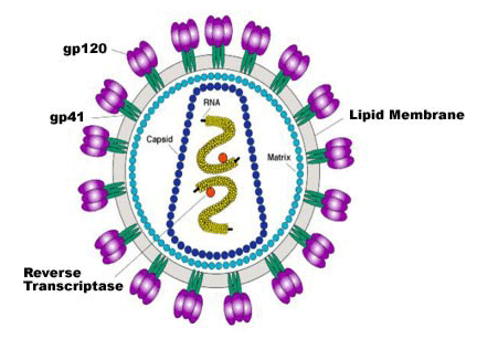
Leukemia

By Applying for disability benefits when you have:

Can I Get Social Security Disability Benefits for Leukemia?

  • How Does the Social Security Administration Decide if I Qualify for Disability Benefits for Leukemia?
  • About Leukemia and Disability
  • Winning Social Security Disability Benefits for Leukemia by Meeting a Listing
  • Residual Functional Capacity Assessment for Leukemia
  • Getting Your Doctor’s Medical Opinion About What You Can Still Do

How Does the Social Security Administration Decide if I Qualify for Disability Benefits for Leukemia?

If you have acute or chronic leukemia, Social Security disability benefits may be available. To determine whether you are disabled by leukemia, the Social Security Administration first considers whether your leukemia is severe enough to meet or equal a listing at Step 3 of the Sequential Evaluation Process. See Winning Social Security Disability Benefits for Leukemia by Meeting a Listing. If you meet or equal a listing because of leukemia, you are considered disabled. If your leukemia is not severe enough to equal or meet a listing, the Social Security Administration must assess your residual functional capacity (RFC) (the work you can still do, despite your leukemia), to determine whether you qualify for benefits at Step 4 and Step 5 of the Sequential Evaluation Process. See Residual Functional Capacity Assessment for Leukemia.
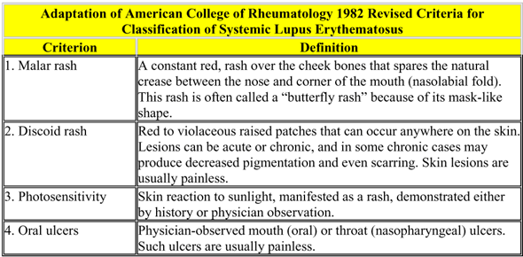
About Leukemia and Disability

What Is Leukemia?

Leukemia is a form of cancer arising in the bone marrow. Through a process known as hematopoiesis, the bone marrow produces the various types of cells that appear in the blood, including red blood cells (RBCs), platelets, and the various types of white blood cells (WBCs) (see Figure 1 below). In leukemia, the bone marrow produces excessive amounts of some type of white blood cell. The WBCs produced cannot carry out their normal functions because they are flawed.

During the process of hematopoiesis, white blood cells begin as blasts. Normally, the blasts then mature into more differentiated forms that carry out specific functions. For example, lymphoblasts eventually turn into mature lymphocytes; myeloblasts become granulocytes. In leukemia, the blasts do not mature properly.

Normal total white cell count in the blood is about 5,000 to 10,000/mm3. In leukemia, the WBC can rise into the 100,000/mm3 range or higher. Red blood cell production is decreased causing (often severe) anemia. Platelets may also be abnormal with high or low counts.

Composition of blood

Figure 1: The composition of blood, including white and red cells and platelets.

Symptoms of Leukemia

Symptoms of leukemia include fatigue and weakness. Patients are often pale from anemia and perhaps bleeding abnormally from some area in the body. Bleeding into the skin produces red-purple spots called petechiae.

Bone pain is common in acute leukemia. Involvement of the skin (leukemia cutis) may be present in a number of forms, such as ulcerations, blisters, raised, flat, or other lesions. Such involvement outside of the blood and bone marrow carries an even worse prognosis than acute leukemia.

Acute Leukemia

All cases of acute leukemia qualify for Social Security disability benefits based on the documented diagnosis. That diagnosis should come from a copy of the formal pathology report about the bone marrow biopsy. See Meeting Social Security Administration Listing 13.06A for Acute Leukemia.

In acute leukemia, the white cells involved are more immature and tend to be more toward the blastic end of development. These leukemias are more dangerous than the chronic forms involving more mature white cells. In adults, acute myelocytic leukemia (AML) is most frequently seen. There are other types of acute leukemia, such as acute monocytic leukemia or acute eosinophilic leukemia that involve different types of white cells. A rare form of hematologic (blood) cancer is hairy cell leukemia, which responds well to chemotherapy.

Over 10,000 new cases of AML are diagnosed yearly in the U.S. and about a third of these patients have a mutation in the FLT3 gene, which in turn produces an abnormal enzyme (tyrosine kinase) that drives the production of leukemic cells. Individuals with the FLT3 gene mutation have only a 10% to 20% cure rate, compared to individuals with other forms of AML who have a cure rate of 40% to 50%. In AML, patients under 60 years of age can expect a remission in 70% to 80% of cases with a 5-year survival of 40% to 50%; older patients only achieve complete remission in about 50% of cases with a 5-year survival of less than 10%.

Experimental drugs known as tyrosine kinase inhibitors are under development, and will probably greatly increase cures while eliminating the toxic side effects of the drugs usually required to treat acute leukemia. A different tyrosine kinase inhibitor used to treat chronic leukemia (Gleevec, imatinib) has been very effective, but is specific for chronic leukemia and will not work for acute leukemia. See Treatment of CML.

Treatment of Acute Leukemia

Chemotherapy is the treatment for acute leukemia, but in some instances a bone marrow or stem cell transplant is done. Some cases are incurable. A state in which there is no evidence of the leukemia after treatment is called remission, but it is not necessarily a cure because a relapse can occur. The longer a remission lasts, the more likely a cure has been achieved. To achieve a complete remission, which is the goal of treatment, there must be no detectable abnormal cells in peripheral blood or bone marrow aspirations.

If bone marrow transplantation is carried out at the time of the first relapse or second chemotherapy-induced remission, a cure can be achieved in about 30% of cases. That figure increases to 50 – 60% if the transplant is done during the first complete remission.

Chronic Leukemia

The chronic leukemias involve proliferation of white cells of a more mature and less cancerous variety than the acute leukemias. Chronic leukemias, while they are extremely serious diseases, do not tend to be quite as severe as the acute forms of leukemia. For that reason, a claimant is not automatically awarded Social Security disability benefits simply based on a diagnosis. See Meeting Social Security Administration Listing 13.06B for Chronic Leukemia.

Some people with chronic leukemia live for years with treatment. For example, some cases of chronic lymphocytic leukemia (CLL), which usually occurs in older people, require no treatment, or only modest treatment. Chronic myelogenous leukemia (CML) causes death in a substantial proportion of people within several years of diagnosis, even with treatment. Advances in treatment, including stem cell or bone transplantation and new drugs, continue to improve the prognosis.

Blastic Transformation of Chronic Leukemia into Acute Leukemia

The danger of a chronic leukemia, especially CML, is that it will change into an acute leukemia—acute myeloblastic leukemia (AML). The change of chronic to acute leukemia is known as a blastic transformation and is associated with an extremely high mortality, usually from infection. A blastic crisis means the majority of granulocyte white blood cells called myelocytes regress to a more immature stage of development in which they are both incapable of their normal specialized functions and are more aggressive as a blood cancer. So, a blastic transformation or crisis is the event most dreaded after diagnosis of CML.

Treatment of CML (Chronic Myelogenous Leukemia)

The transformation of normal myelocytic white blood cells into the cancerous cells of CML occurs by production of an abnormal protein known as BCR-ABL, which enables the cancer cells to live and proliferate. This protein is created by a specific enzyme known as tyrosine kinase.

Several different drugs inhibit tyrosine kinase so that the abnormal BCR-ABL protein cannot be created. Imatinib (Gleevec) is the first drug of its type to target a specific enzyme involved in the leukemic disease process. The side effects of imatinib are infrequent compared to conventional chemotherapeutic agents because it is not broadly toxic to cells like conventional chemotherapy. Nearly 25% of conventional chemotherapy patients will have severe side-effects and they will be much worse than with imatinib.

The overall 5-year survival with imatinib is about 83%, and about 63% remain in major cytogenetic (chromosomal) response after that amount of time. The best predictor for both overall and progression-free survival is a cytogenetic response at one year.

Imatinib can be toxic to the heart in patients with some conditions (hypereosinophlic syndrome and cardiac involvement, or chronic eosinophilic leukemia). These effects are generally controllable with corticosteroids. Older patients who have additional disorders may risk left ventricular dysfunction and congestive heart failure. See Can I Get Social Security Disability Benefits for Congestive (Chronic) Heart Failure? These serious side-effects are unusual; imatinib is usually well-tolerated.

In one study comparing Gleevec to standard chemotherapy with interferon and cytarabine, Gleevec was found to be about 10 times as effective in controlling CML. After 14 months, 68% of patients who received imatinib were completely free of leukemia, compared to only 7% of the interferon-cytarabine group. Furthermore, a much smaller number of patients in the Gleevec group progressed to more serious disease—blastic transformation. Unfortunately, if the patient has already entered a blastic crisis, Gleevec is much less effective, as are other medications. Once a blastic transformation occurs, Gleevec does not improve the very poor survival of standard chemotherapy when used alone. However, far fewer CML patients (1.5%) develop a blastic crisis than would otherwise be the case.

After 5 years of treatment, about 25% of patients taking imatinib discontinue it because of side effects or poor response.

Another tyrosine kinase inhibitor has been developed to be used as a second-line treatment of CML when imatinib fails. Dasatinib (Sprycel) is an oral drug that can be used in all phases of CML, including blastic transformation. A complete hematologic response has been seen with dasatinib in 92% of patients in the chronic phase of CML, and significant improvement in 70% of patients with accelerated CML, CML blast crisis, or Philadelphia chromosome positive acute lymphocytic leukemia (ALL). In one study, 85% of CML patients had achieved a complete hematologic response after 6 months of treatment with dasatinib.

Another tyrosine kinase inhibitor known as nilotinib (Tasigna) was FDA-approved in October 2007 for use in cases resistant to imatinib. A complete hematologic response with no evidence of leukemia has been achieved for at least 6 months using nilotinib. Its long-term effectiveness will require more time to determine. However, nilotinib appears to have more serious and diverse side-effects than imatinib, including thrombocytopenia, neutropenia, and cardiac toxicity, all of which carry a risk of death. Nilotinib can also be used in the accelerated phase of CML in which the disease is progressing with 10-19% blastic forms in peripheral blood. Still, chances of survival with nilotinib are far better than can be expected from untreated CML, which is certain death.

Regardless of the type of treatment, blastic transformation of either CML or CLL is associated with a survival of about 2 to 11 months, depending on the study. Those with lymphoid blastic transformation, as with conversion of chronic lymphocytic leukemia to acute lymphocytic leukemia (CLL to ALL), tend to live a few months longer than those who transform from chronic myelogenous leukemia to acute myelogenous leukemia (CML to AML). The worst prognosis is present when white cells are rapidly progressing to more immature forms and peripheral circulating blood has more than 50% of such blasts.

Continue to Winning Social Security Disability Benefits for Leukemia by Meeting a Listing.
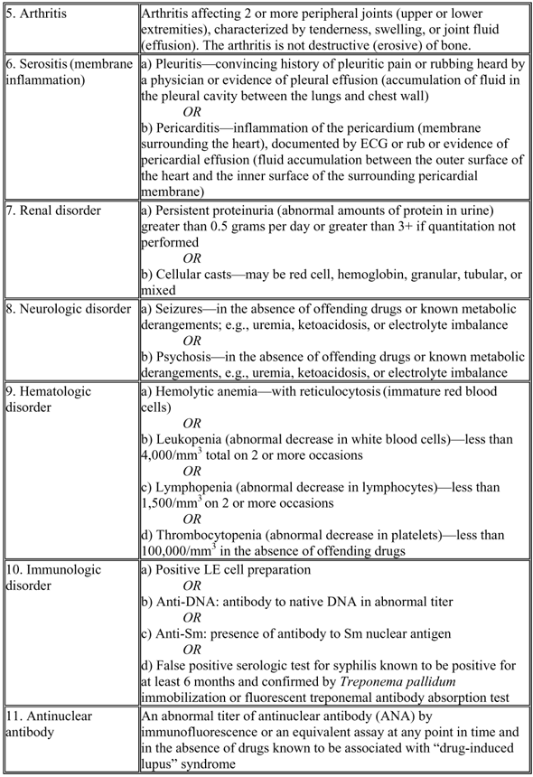
Lupus

By Applying for disability benefits when you have:

Can I Get Social Security Disability Benefits for Lupus?

  • How Does the Social Security Administration Decide if I Qualify for Disability Benefits for Lupus?
  • About Lupus and Disability
  • Winning Social Security Disability Benefits for Lupus by Meeting a Listing
  • Residual Functional Capacity Assessment for Lupus
  • Getting Your Doctor’s Medical Opinion About What You Can Still Do

How Does the Social Security Administration Decide if I Qualify for Disability Benefits for Lupus?

If you have lupus, Social Security disability benefits may be available. To determine whether you are disabled by lupus, the Social Security Administration first considers whether your lupus is severe enough to meet or equal a listing at Step 3 of the Sequential Evaluation Process. See Winning Social Security Disability Benefits for Lupus by Meeting a Listing. If you meet or equal a listing because of lupus, you are considered disabled. If your lupus is not severe enough to equal or meet a listing, the Social Security Administration must assess your residual functional capacity (RFC) (the work you can still do, despite your lupus), to determine whether you qualify for disability benefits at Step 4 and Step 5 of the Sequential Evaluation Process. See Residual Functional Capacity Assessment for Lupus.

About Lupus and Disability

What Is Lupus?

Systemic lupus erythematosus (SLE) is a chronic inflammatory disease that can affect any organ or body system. It is frequently accompanied by severe fatigue, fever, malaise, and weight loss. SLE is much more common in women, who account for 85% to 90% of the cases.

SLE is a multisystem disease. The immune response against the body’s own tissues can affect any organ, with joint, muscle, ocular, respiratory, cardiovascular, digestive, renal, hematologic, skin, neurological, or mental involvement.

Cause of Lupus

Systemic lupus erythematosus (SLE) is an autoimmune disease. Its cause is not well understood, but it does have a genetic component. Numerous “lupus genes” that influence the probability of developing lupus have been identified. SLE probably appears when a person has a particular combination of genes. Due to the complexity of the disease, a cure for SLE is not likely in the near future.

Severity of Lupus

SLE is unpredictable; it is characterized by exacerbations and improvements. It may follow a benign course and be highly responsive to medication, or it may take a sudden severe course leading to early death despite therapy. Any combination of organ systems can be involved in a particular individual, in any degree of severity.

Effects of Lupus

Lupus may result in:

  • Inflammatory arthritis in the joints.
  • Muscle inflammation, pain and weakness.
  • Inflammation of the eye (uveitis), resulting in pain and blurry vision.
  • Respiratory (breathing) problems such as pleuritis, pneumonia, inflammation of the lungs (lupus pneumonitis), and bronchiectasis.
  • Heart problems, such as arrhythmias, murmurs, endocarditis, and cardiomyopathy with heart failure.
  • Digestive problems such as abnormal contractions of the esophagus (dysmotility) or inflammation of arteries (vasculitis) supplying organs of the gastrointestinal system, resulting in pancreatitis, intestinal obstruction, abdominal pain, ulcers, weight loss, or death of intestinal tissue (intestinal infarction) requiring surgical intervention.
  • Kidney problems such as chronic renal failure, which is a common cause of death in SLE.
  • Blood (hematologic) problems which can result in decreased platelets, decreased white cells, or decreased red cells (anemia). Decreased platelets increase susceptibility to bleeding. Decreased white cells increase susceptibility to infection. Anemia results in easy fatigability and weakness.
  • Skin problems leading to scarring, and the need to avoid direct sunlight (photosensitivity).
  • Nervous system involvement resulting in inflammation of the central nervous system—spinal cord and brain.
  • Mental disorders (e.g., psychosis, depression, and organic brain syndrome), which arises from nervous system inflammation.
  • Arterial inflammation (vasculitis) resulting in impaired blood flow to various organs. Impaired blood flow to the hands and feet can decrease tolerance to cold.

Additional possible abnormalities that may be associated with SLE include:

  • Muscle aches (myalgia).
  • Joint pain (arthralgia).
  • Hair loss (alopecia).
  • Fatigue.
  • Fever.
  • Enlarged lymph nodes (lymphadenopathy).
  • Enlarged spleen (splenomegaly).
  • Enlarged liver (hepatomegaly).
  • Sensitivity to cold (Raynaud’s phenomenon).
  • Hypertension.

Diagnostic Criteria Required by the Social Security Administration

There are no universally agreed-upon criteria for making a diagnosis of SLE. The table below shows the diagnostic criteria required by the Social Security Administration.

Criteria for Classification of Systemic Lupus Erythematosus

Treatment for Lupus

Since SLE is incurable, treatment is based on drug therapy that will control symptoms and progression of the disease. Kidney failure is a major cause of death and kidney function must be closely monitored.

Therapy for lupus is based on suppressing the immune system. Systemic corticostroid drugs like prednisone can be highly effective, but their use is limited by potential side-effects (e.g., hypertension, obesity, poor wound healing, osteoporosis, osteonecrosis, cataracts). Methotrexate is another immunosuppressive drug that is useful in treating SLE. The anti-malarial drug plaquenil is often capable of keeping SLE under control.

Specific medications may be required for particular problems like hypertension, depression, and skin lesions.

Every case is different. Some people respond rapidly to maintenance therapy with plaquenil and have minimal symptoms. Others are not so fortunate.

Continue to Winning Social Security Disability Benefits for Lupus by Meeting a Listing.

Multiple sclerosis

By Applying for disability benefits when you have:

Can I Get Social Security Disability Benefits for Multiple Sclerosis?

  • How Does the Social Security Administration Decide if I Qualify for Disability Benefits for Multiple Sclerosis?
  • About Multiple Sclerosis and Disability
  • Winning Social Security Disability Benefits for Multiple Sclerosis by Meeting a Listing
  • Residual Functional Capacity Assessment for Multiple Sclerosis
  • Getting Your Doctor’s Medical Opinion About What You Can Still Do

How Does the Social Security Administration Decide if I Qualify for Disability Benefits for Multiple Sclerosis?

If you have multiple sclerosis (MS), Social Security disability benefits may be available. To determine whether you are disabled by MS, the Social Security Administration first considers whether your MS is severe enough to meet or equal a listing at Step 3 of the Sequential Evaluation Process. See Winning Social Security Disability Benefits for Multiple Sclerosis by Meeting a Listing. If you meet or equal a listing because of your MS, you are considered disabled. If your MS is not severe enough to equal or meet a listing, Social Security Administration must assess your residual functional capacity (RFC) (the work you can still do, despite your MS), to determine whether you qualify for disability benefits at Step 4 and Step 5 of the Sequential Evaluation Process. See Residual Functional Capacity Assessment for Multiple Sclerosis.
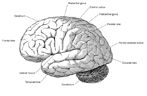
About Multiple Sclerosis and Disability

What Is Multiple Sclerosis?

Multiple sclerosis is a slowly progressive disease of the central nervous system (CNS), i.e., the spinal cord (see Figure 1 below) and brain. The incidence of multiple sclerosis in the U.S. is not reliably known—estimates vary between 250,000 to 350,000 people. In MS a substance known as myelin, which serves as the electrical insulating coating of nerve fiber tracts, is destroyed. Nerve tracts (bundles of communicating fibers) make up the “white matter” of the CNS. Therefore, white matter lesions (wounds or injuries) are characteristic of multiple sclerosis.

Interior view of the spinal cord

Figure 1: Interior view of the spinal cord.

The brain’s white matter lies beneath the brain’s outer cerebral cortex (see Figure 2 below), or “gray matter” that is composed of brain cells (neurons). Most of the volume of the brain is made up of cerebrospinal fluid-filled cavities and white matter. Since the white matter carries information to and from the cortical neurons composing the gray matter, the function of the gray matter can easily be affected by white matter abnormalities. For example, an impulse to move a leg that starts in the motor cortex of the brain could be disrupted when it enters the white matter for transmission to the spinal cord.

Outer cerebral cortex of the brain

Figure 2: Outer cerebral cortex of the brain.

The myelin destruction associated with multiple sclerosis is called demyelinization. MS is not the only disorder that can result in that abnormality. The white matter lesions of demyelinating disorders are called plaques. The plaques of MS occur in multiple and varied locations in the spinal cord and brain, particularly the brain (see Figure 3 below). Brain cells that make myelin are also damaged, but can regenerate to some extent to produce more myelin.

Most people with multiple sclerosis have a normal lifespan, so it is not known as a fatal disorder.

The base of the human brain

Figure 3: Base of the brain.

Cause of Multiple Sclerosis

The cause of MS is unknown. MS is more common in temperate than tropical areas, and moving between geographic regions at certain ages can affect the risk developing the disorder. Theories as to cause have considered a virus, brain trauma, or a problem in the immune system, but these are little more than speculation. Close relatives of people with multiple sclerosis have a higher incidence of the disorder, and this supports the possibility of a genetic contribution.

Genes on multiple chromosomes are suspected of playing some role in MS. People with multiple sclerosis are more likely to have abnormalities in human leukocyte antigens (HLA), proteins coded from chromosome 6 and important in the functioning of the immune system. If there is a genetic contribution to the development of MS, the path of “cause and effect” is not yet understood. There is no genetic test for multiple sclerosis, and even if there is a genetic contribution to cause, unknown environmental factors (such as a particular virus) may also play a role.

Although the primary cause of MS is not known, dysfunction in the immune system is the mechanism by which MS damages nerve tracts. Immune system cells called T lymphocytes (a type of white blood cell) attack the myelin of white matter nerve tracts while B lymphocytes damage the nerve fibers (axons) themselves. While demyelinated areas caused by T cell attack impair nerve impulse transmission, the B cell damage to axons themselves is more serious and slower to repair itself, if repair is possible.

This dual mechanism could explain the different types of MS discussed below. Relapse and remittance of MS may be caused by T cell activity and progression may be caused by B cell activity. All of the answers will have to await future research.

Types of Multiple Sclerosis

Several patterns are possible in the way MS first appears and the way it manifests over time (its clinical course). A typical exacerbation (worsening) of MS lasts 2 to 4 weeks, and takes several months to resolve or improve. However, this pattern is subject to considerable individual variation.

  • Relapsing-remitting MS is the most common type for which Social Security disability benefits are sought. In this form, MS first appears as a series of attacks interspersed with complete or partial improvement (remission), followed by some future relapse.
  • Primary-progressive MS is characterized by a slow progression in clinical severity, although there may be minor temporary stabilization or improvement.
  • Secondary-progressive MS begins as a relapsing-remitting form, then later becomes a primary-progressive form.
  • Progressive-relapsing MS is characterized by worsening, with additional acute exacerbations.

Patients with particularly severe MS might be described as having “malignant MS,” while those with minimal symptoms and limitations for extended periods of time are sometimes referred to as having “benign MS.”

The course of MS can vary significantly from person to person. The Social Security Administration needs detailed medical evidence about how your illness has progressed over time to make the best disability determination.

Diagnostic Categories of Multiple Sclerosis

MS affects women more frequently than men and most often first produces symptoms in the 20 to 40 age range. Disease appearing before age 10 or after age 55 is a strong argument for some other kind of disorder. The diagnosis of MS is still fundamentally clinical—a judgment based on your doctor’s evaluation of the history and physical examination, severity of signs and symptoms over time, and test evidence. The diagnosis cannot be made based on laboratory results alone, and there are no physical abnormalities unique to MS.

Diagnostic categories for multiple sclerosis

Signs and Symptoms of Multiple Sclerosis

There are a large number of possible signs and symptoms of MS. Some patients have no signs or symptoms. The major possibilities are as follows:

  • Muscle weakness sometime associated with muscle atrophy.
  • Spasticity (uncontrolled muscle contractions) – chronic spasticity can impair gait or use of arms; sudden spasms (paroxysmal dystonia) in arms or legs may occur with changes in posture or even certain kinds of sensation.
  • Impairment of pain, temperature, and touch senses – burning, itching, decreased sense of limb position, decreased touch or temperature sensation, etc.
  • Pain (moderate to severe) – sharp pains, including severe facial nerve pain (trigeminal neuralgia).
  • Ataxia – impaired ability to coordinate movement can range from mild to severe.
  • Impaired balance.
  • Tremors.
  • Decreased dexterity – impairment of fine manipulatory ability.
  • Speech disturbances – decreased clarity of articulation (dysarthria) caused by slurring, dyscoordinated speech (speech ataxia), “scanning,” i.e., with unnatural pauses and skipped sounds that result in a staccato-like delivery.
  • Aphasia – difficulty in instigating speech, understanding writing, understanding spoken words, etc.
  • Vision disturbances – paralysis of eye movement and pupillary function (internuclear ophthalmoplegia); double vision (diplopia), blurry vision (optic neuritis), abnormal eye movements (nystagmus); visual field loss is possible but unusual; color vision sensitivity may be decreased, Uhtoff’s phenomenon may occur (visual blurring after exertion or heat exposure).
  • Vertigo – dizziness may be intense, with associated nausea and vomiting; caused by inflammation of the vestibular nerve (vestibular neuronitis).
  • Bladder dysfunction – a common problem that may be urge incontinence, frequency, or decreased bladder sensation resulting in overflow incontinence.
  • Bowel dysfunction – constipation is common; loss of sensation near the anal sphincter may result in bowel incontinence.
  • Sexual dysfunction – frequently present as decreased libido in both sexes; erectile dysfunction in men; decreased vaginal lubrication in women.
  • Depression and anxiety – resulting from poor self-image, sexual dysfunction, worry about spousal rejection, and the debilitating nature of chronic disease.
  • Euphoria.
  • Seizures.
  • Cognitive (rational thinking) abnormalities – present in 40–70% of MS cases but profound mental impairment is uncommon.
  • Fatigue – easy fatigability is associated with both physical and mental activity, and not necessarily associated with obvious physical impairment or depression.
  • Exacerbation with exertion or exposure to heat.
  • Multiple sclerosis may produce findings of both upper and lower motor neuron syndrome, depending on location of the lesions.

Treatment of Signs and Symptoms of MS

Each of the signs and symptoms of MS may require separate treatment. For example, antiepileptic drugs (AEDs) would be required to control seizures and are also helpful in the treatment of the exquisitely painful disorder known as trigeminal neuralgia. Spasticity may be helped by muscle relaxants such as Baclofen (Lioresal), diazepam (Valium) and other drugs. Optic neuritis is treated with corticosteroids like prednisone or intravenous methylprednisolone. Pain may require analgesic or antidepressant medication. A number of different agents have been used to treat fatigue, such as amantadine (Symmetrel), pemoline (Cylert), and antidepressants. Self-injected papaverine in the corpus cavernosum of the penis can help temporarily reverse erectile dysfunction. There are also drugs that can help inhibit an over-active bladder, such as dantrolene (Dantrium).

In addition to drugs, physical therapy, occupational therapy, speech therapy, or psychiatric therapy may be needed in individual cases as appropriate.

Current Therapies for Multiple Sclerosis

Therapeutic Goals

The goals of therapy for MS are threefold: to improve recovery from attacks, to prevent or lessen the number of relapses, and to halt disease progression. Numerous therapies have been tried, but so far, treatment of multiple sclerosis designed to cure or stop disease progression has failed.

Therapies for MS under investigation include various treatments to alter or suppress the immune system, therapies to improve nerve impulse conduction, and therapies to reverse the damage to myelin and oligodendrocytes (the cells that make and maintain myelin in the central nervous system).

Many patients do well with no therapy, especially since many medications have serious side effects and some carry significant risks.

Steroids

Until recently, the principal medications used to treat MS were steroids possessing anti-inflammatory properties; these include adrenocorticotropic hormone (ACTH), prednisone, prednisolone, methylprednisolone, betamethasone, and dexamethasone. While steroids do not affect the course of MS over time, they can reduce the duration and severity of attacks in some patients. The mechanism behind this effect is not known. Because steroids can produce numerous adverse side effects (acne, weight gain, diabetes, seizures, hypertension, cataracts, osteoporosis, psychosis), they are not recommended for long-term use.

Interferons

One of the most promising MS research areas involves naturally occurring antiviral proteins known as interferons. Three forms of beta interferon (Avonex, Betaseron, and Rebif) have been approved by the Food and Drug Administration for treatment of relapsing-remitting MS. Beta interferon has been shown to reduce the number of exacerbations and may slow the progression of physical disability. When attacks do occur, they tend to be shorter and less severe. In addition, MRI scans suggest that beta interferon can decrease myelin destruction. However, if a patient develops neutralizing antibodies to a therapeutic drug then its effectiveness will be reduced. Once neutralizing antibodies have appeared, it does no good to switch to another type of beta interferon since the antibodies associated with the three different drugs cross-react. Common side effects of interferons include fever, chills, sweating, muscle aches, fatigue, depression, and injection site reactions.

Fingolimod

An immunomodulating agent known as fingolimod has much less prominent side effects than interferons and can be taken orally. Fingolimod is aimed at treating relapsing MS, and initial trials show a low relapse rate with continuous oral maintenance, decreased symptoms, and decreased number of lesions on magnetic resonance imaging of the brain. Fingolimod has not been approved by the FDA for human treatment outside of clinical trials. However, there is a good chance fingolimod will turn out to be a useful agent.

Natalizumab

A drug known as natalizumab was approved for treating multiple sclerosis in 2004. Natalizumab decreases the ability of white cells to enter the central nervous system. In 2005, use of the drug had to be suspended due to the risk of a rare but fatal disease known as progressive multifocal leukoencephalopathy (PML). Also, other serious problems such as hypersensitivity reactions can develop. In 2006, the drug was again approved, but only for restricted use in relapsing multiple sclerosis. Patients with progressive MS are not treatable with this drug at the present time. Treatment with natalizumab must be given intravenously on a monthly basis at a special infusion centers.

The significance of a person receiving natalizumab is two-fold for disability adjudication:

  1. The diagnosis must be very secure for the drug to be used; this should suggest to the adjudicator that the claimant has MS beyond any reasonable doubt.
  2. The drug is dangerous; it can be cleared for use only when severe relapsing MS has not responded to other treatments.

Therefore, if you are receiving this drug for treatment of MS, it should be a red flag to the Social Security Administration adjudicator to allow your case when the medical evidence is developed. If the medical evidence does not support an allowance, there is almost certainly a deficiency in the development of that evidence. Even if you have seemingly improved on natalizumab, exceptionally careful consideration of the evidence should be done before concluding that you have achieved a steady-state of improvement that would permit sustained substantial gainful activity.

Continue to Winning Social Security Disability Benefits for Multiple Sclerosis by Meeting a Listing.
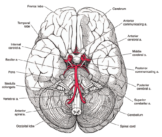
Our Lowell Social Security Lawyer on Applying for Disability While Working

By Applying for disability benefits when you have:, English Blogs, Social Security Disability Library

Our Lowell Social Security Lawyer on Applying for Disability While Working

Lowell Social Security LawyerThe great majority of Social Security disability claimants are not working; although a Lowell Social Security lawyer can explain that there is no prohibition against doing so, it is important to realize that it may be more difficult to be approved for benefits if you continue to work.

Substantial Gainful Activity

This is the dollar amount of monthly income a recipient is permitted to earn while collecting disability. For 2015, the SGA is $1090; however, a Lowell Social Security lawyer cautions that the Social Security Administration can look to other factors in ultimately determining eligibility for disability.

What the SSA Includes and Deducts

Most claimants who are employees will consider their regular gross earnings and compare that amount to the SGA, but the SSA regulations call for a different treatment of certain types of earnings. For instance, bonuses are included but sick pay and vacation pay are deducted to determine your SGA. Additionally, if your impairment requires you to spend money to accommodate your ability to work, those expenses are also deducted. Similarly, if your employer is covering expenses to enable you to work, the SSA should offset that amount in determining your SGA.

Perceived Value vs. Real Value

Some employers actually subsidize an employee who is struggling with an impairment by paying a full salary even though the employee’s true value is less because of his or her physical or mental limitations. The SSA may consider this factor. On the other hand, if you are self-employed, there may be ways to claim you are making less money than is the true value to your business. If so, the SSA may consider your SGA amount to be higher than your declared net profit in the business.

Contact a Lowell Social Security Lawyer for Legal Advice

The rules and regulations governing the disability process are confusing, and more than half of all applicants are initially denied benefits. If you have any questions at any stage of your claim, call the Palma Law Offices, PC, a Lowell Social Security lawyer, at 888-295-4955.

Skip to content